The enhancement and pruning of neural networks occurs most apparently as the baby begins to develop language. Spoken languages can sound very different from each other. In all, human languages produce about 200 different spoken sounds, called phonemes. Spoken English contains just over one-sixth of those possible sounds.
A Japanese-language keyboard suggests some of the potential complexity of learning language.
Brain scans of newborns reveal that in the first few months of life, their brain recognizes the subtle differences in phonemes other than those spoken at home. Japanese infants easily recognize the difference between the sounds made by the letters R and L. However, as the Japanese language has no sound like the letter L, adults raised speaking Japanese lose their ability to distinguish it from the letter R. Similarly, English speakers learning Spanish as adults struggle to separate the subtle sounds of the letters Band P in spoken Spanish.
But babies are able to tell such differences. That’s why it’s far easier to learn a variety of languages as a child. However, as infant brains focus on processing the auditory signals of their native languages, starting at about age 11 months they lose their ability to differentiate some nonnative phonemes. Children and adults who learn new languages after having undergone “phoneme contraction” speak with an accent.
By the time a baby is three or four months of age, its behavior provides clues to its having reached new milestones in brain development. At that age, individual infants differ widely in their reaction to events and in their patterns of brain activity as measured in EEG scans.
Rs & Ls
JAPANESE WHO BEGIN studying the English language as adults struggle with the sound of the letters Rand L. It’s not the tongue that’s to blame-it’s the brain. Newborns can distinguish all phonemes, or language sounds. Between six months and one year of age, however, children lose the ability to process previously unheard language sounds. Their loss is called phoneme contraction. Since the Japanese language slurs Rand L phonemes, adults who are exposed to the separate sounds in English for the first time cannot hear, or articulate, the difference. It’s the same for English speakers learning Japanese. They can learn the words, but it’s too late for the neuronal circuits to get the sounds exactly right.
A pattern of responses known as behavioral inhibition, which includes shyness and fear when exposed to new people and experiences, occurs in one in five healthy four month olds. Their brains show higher levels of electrical activity in the right frontal lobes. Likewise, older babies who cry upon being separated from their mother have more activity in the prefrontal cortex of their right hemisphere than do children who remain calm when mom disappears from sight.
ALBERT & THE RAT
IN A 1913 manifesto, John B. Watson introduced the term behaviorism, which, he wrote, eliminated the “dividing line between man and brute” in asserting that emotions are determined not by DNA but by external stimuli. Watson built on Ivan Pavlov’s foundation of conditioned stimulus response. Foreshadowing the 1932 publication of Aldous Huxley’s novel Brave New World, Watson theorized that “man and brute” alike can be made to order. He guaranteed, for instance, to rear any of 12 random infants to take on the occupation of his choosing. Yet Watson is remembered most, perhaps, for instilling in an infant boyan irrational fear of all things white and furry.
An 11-month-old called Little Albert plays his part in a famous behaviorist experiment.
In 1919, Watson began to work with 11-month-old Little Albert, conditioning him to fear a white rat. To begin with, Albert liked his pet, trying to touch and even hold it. Watson believed this reflected a curiosity innate in all children. Later, a new stimulus was introduced: When Albert reached for the rat, Watson banged a metal bar with a carpenter’s hammer. Albert fell face-forward on the mattress, whimpering. The rat was shown repeatedly, with gong and without, until Little Albert’s congenital fear of loud noises was transferred to the rat. This phobia, Watson later learned, applied also to white rabbits, dogs, a fur coat, and even a Santa Claus mask. Presumably, Watson wrote, Albert could eventually become unconditioned, but the boy was adopted before further experiments could be performed.
Some scientists argue that as the brain incorporates new experiences and makes new connections among neurons, it expresses a form of evolution through the competition of its various neural networks. Nobel Prize-winning neuroscientist Gerald Edelman suggests that the brain’s many networks vie against each other in “neural Darwinism.”
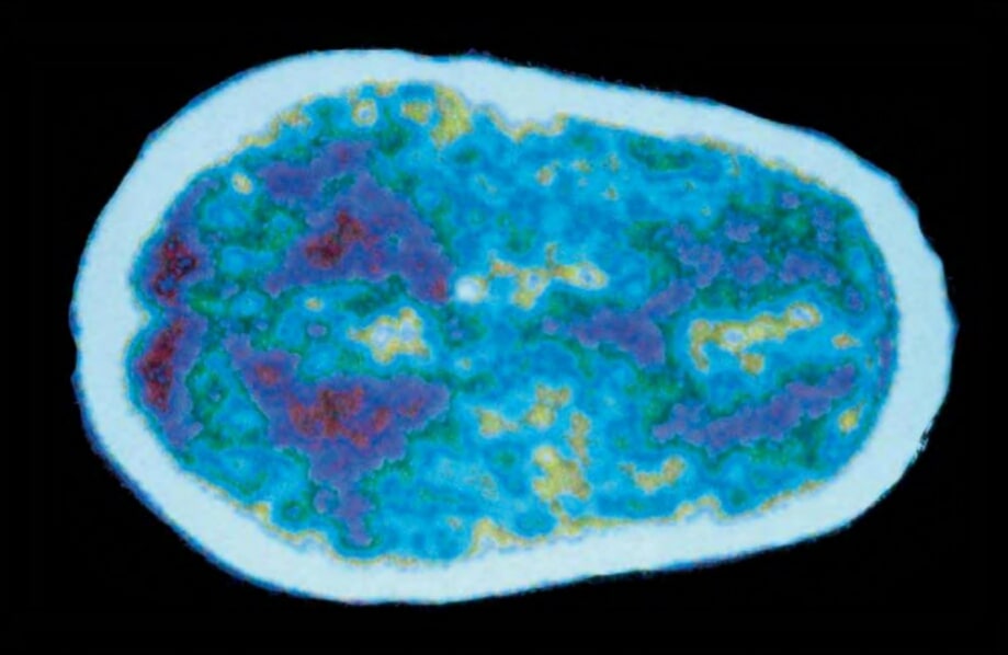
A newborn’s brain (seen above in an MRI) is ready to begin making, remaking, and pruning neural connections by the million.
While genes determine how the brain begins to grow in an embryo, the brain’s extreme complexity and plasticity make it nearly impossible to predict how it will develop in response to a particular stimulus. The complexity of the brain makes it like the weather. Short-term weather forecasts are possible with some degree of confidence, but long-range forecasts become more and more difficult because of the interaction of so many variables. The so-called butterfly effect, which was discovered during computer generated weather simulations in the 1960s, posits that under the right conditions, the flapping of a butterfly’s wings in China can be magnified until it causes a tornado in Texas. As expressed in the brain, a small change in biochemistry under sensitive conditions may have a tremendous impact on the brain’s future development.
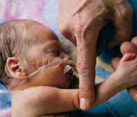
PREMATURE births pose special challenges to the brain. The child emerges from the womb before its neural networks have been established and have gone through initial stages of pruning. Much of the brain development must occur in the buzzing confusion of the world rather than a calm womb, which psychologist Sigmund Freud called the baby’s stimulus barrier. Development of the preemie’s brain occurs without the nutrients and protection of the uterine environment. In addition to difficulties involving regulation of body temperature, digestion of food, and weakened breathing, many preemies suffer brainhemorrhage. Babies who survive amid the chaos of lightsand sounds in a hospital nursery may have their brain overstimulated and may develop problems such as attention disorders and learning disabilities later in life.
Brigham and Women’s Hospital in Boston has attempted to re-create the conditions of the womb in its neonatal intensive care unit. A preemie’s brain reacts with extreme sensitivity to light and loud noises, so the hospital keeps its NICU dark and quiet. Babies get plenty of skin-to-skin contact, to mimic the touch of the womb. They feed on demand. And they’re allowed some freedom of movement, as they would experience inside the womb, rather than being swaddled tightly The result: These babies leave the hospital earlier than those raised in a standard intensive care unit and have an accelerated developmental curve compared with other preemies.
Consider how neural Darwinism finds expression in the early stages of fetal brain growth. Neurons forming from stem cells move through the brain, guided by basic genetic coding. Genes determine how the neurons connect, axon to dendrite, to create the foundation and basic architecture of the brain. However, the precise chemical environment surrounding the newly formed neurons strongly influences how far they migrate and which neighboring neurons they link with. Exposure to substances in the womb, such as alcohol, can disrupt neuronal migration, but there is no guarantee that exposure will or won’t lead to fetal alcohol syndrome. The unpredictability of the complex system that is the human brain makes such precise calculations impossible.
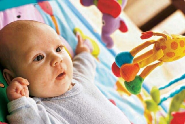
Toys and a mentally stimulating environment help a baby’s brain grow complex neural connections.
Babies don’t learn to walk until about a year after birth, but they are born with the neural program already hardwired.
As people grow older, they take in new experiences. There may be changes in climate, social networks, formal education, and career. To get on in life, people have to adapt to change. Successful adaptation is a matter of rewiring the brain by creating new neuronal connections. Links that promotesurvival and well-being grow stronger. Those that lose their usefulness grow weaker. In a process that resembles natural selection, they lose the competition to stronger neural networks, and they die.
Neural Darwinism provides a new perspective on the brain’s plasticity: As neural networks compete, those that function best get stronger. Changes in the environment encourage changes in the brain by giving new neural networks a chance to flourish. Such evolution of a single brain continues over an entire lifetime.
As a baby emerges from the womb, brain development expands to include processing responses to the baby’s new experiences sights, sounds, smells, actions, sensations, and emotions. Networks of neurons, primed to receive new stimuli,compete for survival. It’s a random battle at first, but soon becomes more organized as environmental stimuli strengthen some connections while others wither. If the baby is exposed to a broad vocabulary and a wide range of music, the connections for language and sound recognition grow stronger. If the baby is kept in an environment lacking in toys and visual stimulation, the baby’s analytical powers may be slow to develop.
ESTABLISHING NETWORKS
Defects in infants’ eyes illustrate the sensitivity of a newborn’s brain and the competing neural networks. When a child is born with a cataract in one eye, that eye is deprived of normal vision, and the portion of the brain that processes information from that eye suffers lack of stimulation. The baby’s one normally functioning eye begins to process all visual information.
NEWBORN SIGHT
WE CAN’T KNOW for certain what the world looks like to a newborn; babies don’t answer interviewers’ questions. However, scientists who study the makeup of new-borns’ eyes and test for whether babies will gaze at objects believe that for the first months of life, children lack the ability to see fine lines and a full spectrum of colors. The world probably looks like a blurred, faded photograph as seen through a card-board tube.
New-borns appear to be hardwired for looking at faces. Shortly after birth, infants will look at faces longer than they will look at any other object.
The “use it or lose it” principle starts to work-with a vengeance. Neural connections develop for the good eye but fail to do so for the eye with the cataract. Unless the cataract is removed shortly after birth, the child will remain blind in that eye. Even if the cataract is removed later, the brain has lost its one chance to develop the neural circuitry to process visual signals from the eye; the eyeball may appear healthy, but it cannot communicate with the brain.
If surgery removes the cataract in time, the strong, already existing neural connections of the stronger eye give it a favored place in brain development. In order to make both eyes work with the same acuity, doctors often patch the stronger eye for a few hours every day. That way, for extended periods, all of the neural development for vision is processed via the weaker eye. Its brain circuitry grows stronger by not having to compete all the time with the good eye.
The process of establishing and strengthening connections in the brain to process vision underscores the fact that certain periods are absolutely critical to proper functional development. While the brain retains a measure of plasticity among existing networks, it also seldom offers a second chance for establishing those networks at an early age. In other words, the brain cannot expand and reconnect a neural network that doesn’t exist or one that exists, like a dead-end road, without functional traffic.
The first, and easiest, thing a mother to be can do is to eat for two: This doesn’t mean doubling up on servings it means remembering that the vitamins and minerals from a well-balanced diet not only nourish mom’s brain and body but the brain and body of her developing baby. Pregnant women need proper amounts of folic acid, vitamin B12 (crucial to the functioning of the central nervous system), fatty acids, iron, and other nutrients. She should consult her obstetrician about taking prenatal vitamins, which contain many of these substances and fill in any nutritional gaps in her diet.
Getting plenty of exercise is important to both the mother and her developing baby.
Good nutrition is vital for healthy brain development. Lack of nutrients at crucial moments in fetal brain development leads to a drop or even a halt in the creation of neurons. Babies born after suffering malnutrition often display a smaller brain and have cognitive disabilities. Lack of folic acid (found abundantly in bread, beans, pasta, spinach, and orange juice) raises the chances of a child being born with spina bifida. On the other hand, too much of a good thing can be bad. Overabundance of certain vitamins, including A and D, can cause toxic reactions in the fetal brain. The best advice for a mother to be is to consult her doctor about the best diet for her, one with lots of fresh fruits, leafY green vegetables, legumes, whole grains, and lean meats.
AVOID ALCOHOL
To decrease the chances of neurological defects, moms to be should also avoid many substances that can harm an unborn child’s brain, such as alcohol. In 1899, William Sullivan, a doctor who studied babies born in an English women’s prison, discovered much higher rates of still-births among mothers who drank heavily. He suspected a link between alcohol and fetal health when he noted that mothers who gave birth to babies with severe birth defects in the outside world had healthy babies in prison, where they were denied alcohol.
It would take more than seven decades before researchers at the University of Washington cataloged the recurring patterns of birth defects as fetal alcohol syndrome. When pregnant women drink heavily, their children are at high risk of having a malformed heart and limbs, a smaller brain, reading and math disabilities, hyperactivity, depression, and distinctive facial abnormalities. Mental retardation also is possible. Unfortunately, alcohol’s most devastating impact on a developing fetus occurs early in the pregnancy, when the mother may not even know she is carrying a child. And small amounts in the first trimester cause more damage than greater alcohol consumption later on, apparently because of alcohol’s impact on the migration of developing neurons In the fetal brain. Normally, neurons stop their travels when they reach their intended destinations. The presence of alcohol makes them overshoot and die.
JUST SAY NO
Other substances harmful to adults are even more so to a developing fetus, whose brain is especially sensitive to its chemical environment. Tobacco, illegal drugs such as cocaine, and environmental toxins, all of which do some level of harm to an adult’s body, deliver hammer blows to a developing fetus and can even cause harmful impacts on sperm cells, so men should consider their levels of exposure before trying to start a family. Sperm live for about three months. To minimize the chances of their sperm being adversely affected by alcohol, tobacco, drugs, and toxins, fathers to be should avoid exposure to such harmful substances for 90 days.
Drugs taken by pregnant women can cause abnormalities in the developing fetus.
For pregnant women, tobacco smoke is the most common environmental hazard to a fetus. Nicotine in tobacco causes blood vessels to constrict; an affected fetus gets less blood, and its heart rate decreases. Furthermore, nicotine becomes more concentrated in the fetus’s body than in that of the mother. Like alcohol, nicotine is believed to interfere with neuronal migration, connection, and development. Spontaneous abortion rates nearly double for mothers who smoke. Babies carried to term are more likely to be mentally retarded and have congenital abnormalities.
SEEK OUT HIDDEN RISKS
Toxins harmful to a fetus range from obvious hazards such as the poisons in pesticides to common and seemingly harmless substances such as vitamin A, which in high concentrations (such as in acne medication) harms a fetus’s brain. Lead particles, many over the counter and prescription medicines, x-rays, and some cancer drugs also poison a developing brain.
The jury is out on the possible impact of antidepressants. A pregnant woman’s use of Prozac, a common prescription only treatment for depression, so far has been shown to have no impact on her child’s
Migrating neurons are helped along by glial cells. They support and nourish the neurons on their journeys. Some help regulate the neurons’ metabolism, and others coat the nerve cells’ axons with myelin, a fatty substance that provides electrical insulation and thus controls the speed of communication along neural networks.
Although the brain of a fetus at about eight months after conception weighs only a pound, or about a third of an adult’s, it contains twice as many neurons. Chemical signals called trophic factors influence how individual neurons connect to each other, but the survival of those connections depends on repeated communication across the synapses.
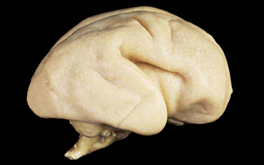
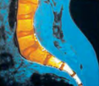
A fetal brain at 24 weeks, with spinal cord at left, has yet to develop characteristic cerebral folding
The brain cannot possibly sustain biochemical reactions across all of its neural connections, and so the weakest connections begin to die, through a process known as pruning. In the last stages of fetal development in the womb, about half of all neurons die. The loss is normal; it eliminates many of the connections that are weak or improper for efficient brain function, leaving behind the strongest and fittest neurons.
FIRST DESCRIBED 4,000 years ago, spina bifida is a malformation of the fetal spinal column that has been linked to a diet deficient in folate, a B vitamin, in pregnant women.
From the Latin for “spine split in two,” the birth defect occurs in 1 to 2 births per 1,000. One or more vertebrae, particularly in the small of the back, don’t grow the bony projections called vertebral arches that point away from the center of the body. Often a cyst bulges outward from the spine, encompassing spinal tissues, cerebrospinal fluid and even parts of the cord itself. Large cysts likely signal severe neurologicalimpairment; a portion of the body’s central nervous system, designed to be safely protected from the outside world behind walls of tissue and bone, lies exposed. When the spinal cord is so compromised as to lose function, the infant may suffer paralysis of the legs and bladder, as well as bowel incontinence.
As a preventative measure, since 1998 all bread, pasta, and flour produced In America contains supplemental amounts of folate. The vitamin, found in green, leafy vegetables, helps the body grow new cells, but how its lack can trigger the disorder remains unclear. Genetics playa role, as the highest incidencerates occur among the citizens of Ireland and Wales as well as their immigrant descendants.
Surgery often can close openings over the exposed portion of a spine and reconstruct misshapen vertebrae, but many impairments remain for a lifetime.
The most dynamic growth occurs in the cerebral cortex, the largest and outermost layer of the brain.During the first months of fetal development, when 250,000 new nerve cells are being created every minute, neurons begin to take on specialized tasks.
First, they inch their way from where they were formed by cell division to their permanent home in other regions of the brain. Most go toward the cortex, but some move into the cerebellum and other portions of the brain. This process, known as migration, is quite remarkable for the distance the neurons must travel as well as their ability to navigate surely along the tangled pathways of the developing brain. Millions of neurons migrate a distance equivalent to a person hiking from Los Angeles to Boston. Amazingly, they manage to arrive at Paul Revere’s house, the U.S.S. Constitution, or Faneuil Hall without ever consulting a map.
Once the migrating neurons reach their destination, they developed axons and dendrites to reach out and make connections with other neurons. Like roads that connect to create a grid for traffic, neurons set up systems of communication that link all parts of the brain. Some pathways receive huge amounts of sensory traffic and become the equivalent of information highways. Others turn into dead ends or decay into crumbling blacktop from lack of use.
You can’t clone a brain. And even if you could, it wouldn’t turn out like the original. Sensitivity to initial conditions in the womb coupled with differences in environment after birth would significantly alter development despite the identical genetic code.
UNDERSTANDING MIGRATION
The brain reacts with extreme sensitivity to anything that influence neuronal migration. Only a few decades ago, neuroscientists believed that each neuron had its own special, predetermined location when it set out on its trek across the brain. Now, researchers have found that neurons take on different characteristics because of their journey and their destination. To take just one example, neurons that process oral communication are not inherently preprogrammed to be speech neurons. Instead, they become speech neurons by migrating to the areas of the brain associated with language.
This discovery prompted new understanding of a wide variety of brain disorders. If something interferes with neurons migrating to their intended destinations and not overshooting or undershooting their targets the results can be powerful. Such disorders as autism, schizophrenia, dyslexia, and epilepsy have been at least partly linked with abnormalities in neuronal migration.
Fetal alcohol syndrome has also been linked to problems in migration. The brain’s hypersensitivity to toxins that impede migration underscores the warnings given to expectant mothers to avoid exposing a developing baby to alcohol, tobacco smoke, drugs, or other chemicals that may interfere with healthy brain development.


![NEWBORN NEURONS [ BRAIN ]](https://humanityuapd.com/wp-content/uploads/2022/12/20221206_164529_0000-1024x1024.png)
![A GOOD START FOR THE PREGNANT MOTHER - DIET, PREVENTION, RISK [ Getting plenty of exercise is important to both the mother and her developing baby. ]](https://humanityuapd.com/wp-content/uploads/2022/12/Screenshot_2022-12-04-18-51-25-523_com.google.android.apps_.docs2_.png)
![A GOOD START FOR THE PREGNANT MOTHER - DIET, PREVENTION, RISK [ Drugs taken by pregnant women can cause abnormalities in the developing fetus. ]](https://humanityuapd.com/wp-content/uploads/2022/12/Screenshot_2022-12-04-18-52-01-283_com.google.android.apps_.docs2_.png)


You must be logged in to post a comment.