The enhancement and pruning of neural networks occurs most apparently as the baby begins to develop language. Spoken languages can sound very different from each other. In all, human languages produce about 200 different spoken sounds, called phonemes. Spoken English contains just over one-sixth of those possible sounds.
A Japanese-language keyboard suggests some of the potential complexity of learning language.
Brain scans of newborns reveal that in the first few months of life, their brain recognizes the subtle differences in phonemes other than those spoken at home. Japanese infants easily recognize the difference between the sounds made by the letters R and L. However, as the Japanese language has no sound like the letter L, adults raised speaking Japanese lose their ability to distinguish it from the letter R. Similarly, English speakers learning Spanish as adults struggle to separate the subtle sounds of the letters Band P in spoken Spanish.
But babies are able to tell such differences. That’s why it’s far easier to learn a variety of languages as a child. However, as infant brains focus on processing the auditory signals of their native languages, starting at about age 11 months they lose their ability to differentiate some nonnative phonemes. Children and adults who learn new languages after having undergone “phoneme contraction” speak with an accent.
Some scientists argue that as the brain incorporates new experiences and makes new connections among neurons, it expresses a form of evolution through the competition of its various neural networks. Nobel Prize-winning neuroscientist Gerald Edelman suggests that the brain’s many networks vie against each other in “neural Darwinism.”
A newborn’s brain (seen above in an MRI) is ready to begin making, remaking, and pruning neural connections by the million.
While genes determine how the brain begins to grow in an embryo, the brain’s extreme complexity and plasticity make it nearly impossible to predict how it will develop in response to a particular stimulus. The complexity of the brain makes it like the weather. Short-term weather forecasts are possible with some degree of confidence, but long-range forecasts become more and more difficult because of the interaction of so many variables. The so-called butterfly effect, which was discovered during computer generated weather simulations in the 1960s, posits that under the right conditions, the flapping of a butterfly’s wings in China can be magnified until it causes a tornado in Texas. As expressed in the brain, a small change in biochemistry under sensitive conditions may have a tremendous impact on the brain’s future development.
PREMATURE births pose special challenges to the brain. The child emerges from the womb before its neural networks have been established and have gone through initial stages of pruning. Much of the brain development must occur in the buzzing confusion of the world rather than a calm womb, which psychologist Sigmund Freud called the baby’s stimulus barrier. Development of the preemie’s brain occurs without the nutrients and protection of the uterine environment. In addition to difficulties involving regulation of body temperature, digestion of food, and weakened breathing, many preemies suffer brainhemorrhage. Babies who survive amid the chaos of lightsand sounds in a hospital nursery may have their brain overstimulated and may develop problems such as attention disorders and learning disabilities later in life.
Brigham and Women’s Hospital in Boston has attempted to re-create the conditions of the womb in its neonatal intensive care unit. A preemie’s brain reacts with extreme sensitivity to light and loud noises, so the hospital keeps its NICU dark and quiet. Babies get plenty of skin-to-skin contact, to mimic the touch of the womb. They feed on demand. And they’re allowed some freedom of movement, as they would experience inside the womb, rather than being swaddled tightly The result: These babies leave the hospital earlier than those raised in a standard intensive care unit and have an accelerated developmental curve compared with other preemies.
Consider how neural Darwinism finds expression in the early stages of fetal brain growth. Neurons forming from stem cells move through the brain, guided by basic genetic coding. Genes determine how the neurons connect, axon to dendrite, to create the foundation and basic architecture of the brain. However, the precise chemical environment surrounding the newly formed neurons strongly influences how far they migrate and which neighboring neurons they link with. Exposure to substances in the womb, such as alcohol, can disrupt neuronal migration, but there is no guarantee that exposure will or won’t lead to fetal alcohol syndrome. The unpredictability of the complex system that is the human brain makes such precise calculations impossible.
Toys and a mentally stimulating environment help a baby’s brain grow complex neural connections.
Babies don’t learn to walk until about a year after birth, but they are born with the neural program already hardwired.
As people grow older, they take in new experiences. There may be changes in climate, social networks, formal education, and career. To get on in life, people have to adapt to change. Successful adaptation is a matter of rewiring the brain by creating new neuronal connections. Links that promotesurvival and well-being grow stronger. Those that lose their usefulness grow weaker. In a process that resembles natural selection, they lose the competition to stronger neural networks, and they die.
Neural Darwinism provides a new perspective on the brain’s plasticity: As neural networks compete, those that function best get stronger. Changes in the environment encourage changes in the brain by giving new neural networks a chance to flourish. Such evolution of a single brain continues over an entire lifetime.
The most dynamic growth occurs in the cerebral cortex, the largest and outermost layer of the brain.During the first months of fetal development, when 250,000 new nerve cells are being created every minute, neurons begin to take on specialized tasks.
First, they inch their way from where they were formed by cell division to their permanent home in other regions of the brain. Most go toward the cortex, but some move into the cerebellum and other portions of the brain. This process, known as migration, is quite remarkable for the distance the neurons must travel as well as their ability to navigate surely along the tangled pathways of the developing brain. Millions of neurons migrate a distance equivalent to a person hiking from Los Angeles to Boston. Amazingly, they manage to arrive at Paul Revere’s house, the U.S.S. Constitution, or Faneuil Hall without ever consulting a map.
Once the migrating neurons reach their destination, they developed axons and dendrites to reach out and make connections with other neurons. Like roads that connect to create a grid for traffic, neurons set up systems of communication that link all parts of the brain. Some pathways receive huge amounts of sensory traffic and become the equivalent of information highways. Others turn into dead ends or decay into crumbling blacktop from lack of use.
You can’t clone a brain. And even if you could, it wouldn’t turn out like the original. Sensitivity to initial conditions in the womb coupled with differences in environment after birth would significantly alter development despite the identical genetic code.
UNDERSTANDING MIGRATION
The brain reacts with extreme sensitivity to anything that influence neuronal migration. Only a few decades ago, neuroscientists believed that each neuron had its own special, predetermined location when it set out on its trek across the brain. Now, researchers have found that neurons take on different characteristics because of their journey and their destination. To take just one example, neurons that process oral communication are not inherently preprogrammed to be speech neurons. Instead, they become speech neurons by migrating to the areas of the brain associated with language.
This discovery prompted new understanding of a wide variety of brain disorders. If something interferes with neurons migrating to their intended destinations and not overshooting or undershooting their targets the results can be powerful. Such disorders as autism, schizophrenia, dyslexia, and epilepsy have been at least partly linked with abnormalities in neuronal migration.
Fetal alcohol syndrome has also been linked to problems in migration. The brain’s hypersensitivity to toxins that impede migration underscores the warnings given to expectant mothers to avoid exposing a developing baby to alcohol, tobacco smoke, drugs, or other chemicals that may interfere with healthy brain development.
WHEN SPERM meets egg, the merger of a father’s and mother’s DNA triggers the start of a new life. Encoded in the tens of thousands of genes that make up a human being are a substantial fraction that will create the brain and central nervous system. You won’t find the child’s personality, emotions, and ideas buried in the code; they arise instead as the brain develops and interacts with its environment after birth. Nevertheless, the explosion of cell development that begins with conception is the first step toward forming the brain and all of the hopes and dreams it will one day contain.
As an embryo develops into a fetus, the brain grows and differentiates rapidly.
DIVISIONS & LAYERS
In its first phases of development, the fertilized egg, or zygote, undergoes a rapid series of divisions. One cell becomes two, two become four, four become eight, and so on until the exponential divisions Create a tiny, hollow ball of hundreds of cells nearly uniform in design.Two weeks after conception, the sphere of cells, still dividing, takes the first step in the series of physical changes to construct a differentiated body and begin the process of becoming human.
First, a dent appears in the sphere. Cells move into the indentation, which folds under the surface of the sphere. The folding creates three layers of cells: an outer layer called the ectoderm, an inner layer called the endoderm, and a middle layer called the mesoderm. In the following weeks, these three layers grow into the tissues that give rise to the body’s major systems: Endoderm becomes digestive tract; mesoderm creates muscles, skeleton, heart, and genitalia; and ectoderm forms brain, spine, nerves, and skin.
Lots of gentle handling produced increased serotonin, a neurotransmitter that dampens aggression, in baby rats. Grown into adults, the rats lived longer and handled stress better.
BUDDING BRAIN
The nascent brain makes its first appearance at about four weeks after conception, when a thin, spoon-shaped layer of cells called a neural plate emerges at the head end of the embryo. Major characteristics of the future brain already are in place just one month into fetal development. Hemispheres later will develop on either side of a groove down the center of the neural plate, creating the bilateral symmetry of the human brain.
As the fetus grows, the bowl of the spoon will become the brain itself, while its handle grows into the spinal cord. And as the neural plate folds to form a tube, swellings in the original spoon shape become the forebrain, midbrain, and hind brain. As they develop, they work together to form the major sections of the brain, from the cerebrum at the top of the head to the thalamus, hypothalamus, cerebellum, and spinal cord at the back and lower end.
As modern humans evolved from their hominid ancestors, their brain development continued with increasing specialization of regions and functions. One hypothesis suggests that the differences between the left and right hemispheres of the human brain can be traced tohumans’ simian ancestors swinging through trees. Grasping one limb after another requires the arms to act independently instead of in unison. Perhaps the ancestors of humans began emphasizing the use of one arm over another, encouraging greater neuronal development in the hemisphere that controlled action on that side of the body.
One of the most pronounced differences between brain hemispheres can be observed in dissection of cadavers. The brain region mainly responsible for speech, the planum temporale, is larger in the left hemisphere of two-thirds of human brains. The left-handed nature of language is evident across time and stage of life. Full-term fetuses exhibit larger, speech-related regions in the left hemisphere than in mirror locations on the right hemisphere. The same was true of Neanderthals, according to the telltale marks on the inside of their 50,OOO-year-old skulls made by contact with their gyri and sulci.
GENDER DIFFERENCES
The two sexes also experience differences in brain function. Men are more likely to be left-handed, dyslexic, hyperactive, and autistic. Women are more likely to suffer migraines and, on average, have weaker spatial functioning. Women, though, generally outperform men in the fine motor skills of their fingers, and they learn to speak their native language earlier and foreign languages more easily than men. The bottom line, however, is that if you were to look at two brains on a laboratory table-one from a man, and the other from a woman-you probably wouldn’t be able to tell any difference.
In men, the third interstitial nucleus of the hypothalamus typically is twice as big as it is in women’s brains. The hypothalamus is crucial to sexual behavior, as well as regulation of body temperature, eating, and drinking. Furthermore, women’s and men’s brains differ in response to orgasm. PET scans show less activity in a woman’s prefrontal cortex and in a man’s amygdala during sexual climax, while both sexes experience more neuronal firing in the cerebellum.
GENDERED BRAIN
THE SEXES DIFFER in cognitive ways. A big one involves spatial orientation. Men typically use mental maps, while women prefer landmarks. Men would likely give directions by saying, “Drive north 2.2 miles, turn east, and drive 1.5 miles,” whereas women would more likely say, “Drive toward the mountains until you see the barn, turn right, and go to the pond.” Small wonder that one sex may get frustrated giving directions to the other. Women take the prize for remembering objects’ locations-where are those keys?- while men win at abstract spatial reasoning, such as mentally rotating objects. As a group, men have a wider dispersal of scores on some mental tests.
PREPROGRAMMING
Much human behavior arises from culture and environment. Some, however, appears to be prewired into the brain. The capacity for language appears to be so strongly encoded that children raised without exposure to any language will make up their own.
Communication is an evolutionary favored social activity that helps humans compete with other animals for resources necessary for life. Similarly, the brain’s ability to process and integrate visual stimuli exists almost immediately after birth. At only a few weeks old, an infant raises its arms to protect itself from the approach of an object. Sight, texture, and size appear to be aspects of object recognition that the brain is prewired to bring together for self-defense.
Some of humanity’s evolutionary history can be observed in the development of a human fetus. As chicken and human embryos develop, for example, they experience a stage where they both have a tail, as well as arches and slits in their neck remarkably like the gill slits and arches found in fish. Thus, scientists in the late 20th century concluded that chickens and humans most likely shared a fish-like ancestor, based not only on visual evidence but also on DNA and fossil records. Not all ancestral characteristics become evident during fetal development, but enough similarities exist to suggest an evolutionary thread.
A few days after conception, a human embryo’s cells begin to specialize. Some form a simple neural plate, which changes into a groove and then a tube. The huge cerebral cortex that distinguishes the human brain develops last, in the final months before birth, just as it evolved from humanity’s simian ancestors two million years ago relatively late on the evolutionary tree. Like an hour-long film compressed into a few seconds, the pageant of growth and diversity in the fetal brain roughly condenses a half billion years of animal evolution into nine months of flesh and blood transformation.
The common animal ancestors of humans and other animals are suggested by common elements of animal brains. The more complex structures of the late developers overlie the simpler forms of creatures that evolved earlier, and thus lower on the evolutionary tree.
PAVLOV’S DOGS
AT FIRST; Russian physiologist Ivan Pavlov (1849-1936) wanted only to know the neural link between dinner and dog drool. To find out, he anesthetized his test subject and detached its salivary duct, lightly stitching this to the dog’s outer cheek. Then, placing food in the dog’s mouth, he could eaSily collect and calculate its salivary response. In this way he hoped to unlock the mysteries of the canine nervous system.
After repeated experiments, Unfortunately the dog seemed to catch on and began to salivate before the food had arrived. Clearly this was a problem. How could Pavlov understand salivary response to food in the mouth if the response occurred in the absence of food? Initially puzzled, Pavlov realized he’d stumbled upon something even more intriguing than his original objective. As environmental factors determine evolutionary adaptations within a species, he concluded, so too must external forces mold the behavior of an individual.
Ivan Pavlov observes one of the dogs he subjected to conditioned behavior experiments.
From a knee-jerk defense mechanism to the performing of Rachmaninoff, acquired reflexes are the building blocks of learning. And if dogs’ brains were sophisticated enough to make such connections, imagine what human brains could do.
Pavlov soon discovered he could condition animals to respond to arbitrary stimuli. If a snack was repeatedly paired with buzzer, whistle, or A-minor triad on the piano-he rarely used that legendary bell-the dog would begin to salivate at sound alone. But a slight variation-B-flat minor, perhaps or A minor in a different octave-triggered no response. The same held for shapes, clocks, shades of gray,melodic patterns, light and rotating objects.
EVOLUTION – GROWTH & ADAPTATION OF THE HUMAN BRAIN [ BRAIN DEVELOPMENT ]
FROM THE single celled product of conception, the human animal grows into a complex, uniquely cognitive being. Evolution has built upon older, more primitive animal brain forms to lead humanity to emotion and rational thought. Over eons of time, neural circuitry has developed to promote and continue to promote individual and collective survival. That’s because the human brain is “plastic,” primed from an extremely young age to learn and change.
A six-month-old girl examines her reflection. From birth, humans appear to be drawn toward faces.
EVOLUTION
THE DEVELOPMENT of the human brain is written in millions of years of evolution, its story still unfolding.
Neurons began to emerge with the appearance of multicellular animals. The earliest neural connections formed primitive networks of cells in tiny life-forms swimming in primordial oceans. Today, such systems can still be found in simple life-forms such as jellyfish.
SIMPLE BRAINS
Animals with only the barest collection of neurons can function with surprising sophistication. The marine snail Aplysia has only about 2,000 neurons, yet it is capable of movement, reaction to touch, sensation, and all of the things that make a snail live like a snail. It even can learn despite lacking a true brain. Aplysia’s neurons organize themselves into clumps called ganglia at various points on its tiny body, creating a maze of connections. These neural clumps can amplifY or tamp down electrochemical signals as they pass from neuron to neuron; its neural connections can be strengthened or weakened just as in human brains. Scientists have found that when they shock Aplysia’s tail, it reacts by reflexits neural network contracts the affected flesh to pull it away from the source of the shock. However, things get interesting when the shock is preceded by a light touch against the snail’s flesh. After a few repetitions, the lowly Aplysia has enough neural complexity to connect the two sensa- tions: touch, followed by pain. In time, the light touch alone, with no electric shock afterward, is enough to make the snail recoil as if in pain.
An octopus’s brain is dime size, but it can solve simple problems such as moving barriers to get food.
CHARLES DARWIN KNEW he had opened a tinderbox when he published On the Origin of Species in 1859. He laid out a theory of evolution through natural selection: Individuals that have a biological advantage are more likely to outlive their peers and pass their edge to offspring. A gazelle that is a bit faster than another may outrun the lion and breed fast children the next day. Cuidado, Darwin wrote in his notebook, using the Spanish for “careful.” Taken toits logical conclusion, even humans fell under his theory-an idea Darwin down-played at first because he knew it would be unpopular.
When the nervous and endocrine systems get out of balance, the resulting dearth or overabundance of hormones can cause havoc. Consider just one hormone. The pituitary gland in the brain stores antidiuretic hormone (ADH), also called vasopressin, which is created by the hypothalamus. ADH helps regulate the body’s water content through its ability to prevent the formation of urine, which contains water expelled by cells.
Neurons in the hypothalamus monitor the water content of the blood and call for the release or withholding of ADH when the blood contains too much or too little water. The dry mouth you experience on the morning of January 1 may be a result of too much partying the night before; excessive alcohol consumption suppresses the release of ADH, causing excessive urination and thus dehydration and cotton mouth.
Blueberries are rich in acetylcholine and antioxidants, making them an excellent food for brain health.
When the hypothalamus and pituitary fail to regularly create and release enough ADH, often through damage to the hypothalamus or the pituitary, the result is diabetes insipidus. Patients with this disorder urinate frequently and are constantly thirsty. Mild forms of diabetes insipidus can be treated simply: As long as the brain’s ability to recognize thirst is undamaged, patients can compensate for dehydration by drinking plenty of water whenever they feel the need.
DIABETES MELLITUS
Diabetes mellitus creates a lack of the hormone insulin, resulting in heavy losses of blood sugar through urination. Insulin arises in the pancreas, a gland that produces enzymes important for digestion. Insulin’s influence is most apparent just after a meal, as it works to take glucose out of the bloodstream to use it for energy in the body’s cells. Insulin also helps store fat and synthesize proteins.
Diabetes mellitus occurs when the pancreas doesn’t produce enough insulin. Its lack leads to excess blood sugar levels, resulting in dehydration through urination, fatigue, weight loss, nausea, abdominal pain, as well as extreme thirst and hunger. The most common treatment is for the afflicted to test their blood sugar levels and inject themselves with insulin when needed. Accidental overdoses are the most common cause of hypoglycemia, which occurs when too much insulin in the bloodstream lowers blood sugar dangerously. Eating a piece of candy or sipping a glass of orange juice helps restore sugar levels.
Regular tests help diabetics monitor levels of glucose in the bloodstream.
CLASSIFICATIONS
Diabetes formerly was classified into “juvenile onset” and “adult onset” varieties because of the typical time frame for diagnoses-ages eight to twelve in children, and forty to sixty in adults. The classification system changed when doctors analyzed symptoms that did not match up well with ages. Patients whose body produced no insulin at all were reclassified as “insulin dependent,” while those whose body made insufficient amounts became “non insulin dependent.” The former now is called Type 1, and the latter Type 2.
Type 1 diabetes IS commonly diagnosed in children, teens, and young adults. Symptoms usually come in a rush, shortly after the patient s Immune system turns on itself and destroys the insulin-producing cells of the pancreas. Lack of insulin used to be a death sentence. Now patients survive with regular injections of insulin, either by syringe or an automatic pump and catheter.
Diabetes mellitus gets its modern name from the Greek for “overflow” (diabetes) and the Latin for “honey” ( mellitus). Overflow is a reference to the symptom of frequent urination, and honey refers to the glucose that appears in the urine. Ancient physicians would diagnose the condition by tasting urine for sweetness.
Type 2 is the more common variety and can begin at any age. It usually starts because the body’s liver, fat, and muscle cells fail to use insulin efficiently. That causes glucose levels to rise in the blood- stream. Feedback mechanisms in the peripheral nervous system detect the increase and trigger production and release of more insulin in the pancreas to offset the higher glucose levels and maintain homeostasis. However, the pancreas cannot keep up the extra production forever. Diet, exercise, weight loss, and medication are common methods of keeping Type 2 diabetes in check.
MAPPING SEIZURES [ DELICATE BALANCE – THE NERVOUS SYSTEM ]
Seizures may occur in any part of the brain; their point of origin often can be mapped. Some occur as a result of lesions in specific domains. Nineteenth-century doctor John Hughlings Jackson, an aloof but meticulous researcher, posited that lesions would produce two effects. He based this belief on the idea that most of the neurotransmitters in the brain at any given moment inhibit action. A minority of neurons at anyone time release neurotransmitters that bind to receptors. Others do nothing. Thus, Jackson said lesions would produce negative reactions because of the destruction of brain tissue. However, they also would have the opposite reaction of freeing other, healthy areas of the brain, which previously had been suppressed.
The minus and plus aspects of brain damage appeared to match the observed effects of a brain tumor in a teenage girl named Bhagawhandi in the 1970s. A neuroscientist who observed the girl diagnosed a malignant brain tumor. As the tumor grew to press on her temporal lobe and her brain started to swell, she suffered a series of seizures. They grew more frequent. However, whereas her initial seizures were intense grand mal convulsions, her new manifestations, localized in the temporal lobe, were weaker. She began experiencing dreamy states in which she saw visions of her home in India. Far from being unpleasant, they made her happy-“They take me back home,” she said. She remained peaceful and lucid during her episodes. The seizures killed her in a few weeks, but doctors often noted the rapt expression on her face as she moved deeper into her visions. Only a few diseases of the central nervous system produce pleasure. Anything that pushes the brain out of homeostasis is more likely to bring pain and discomfort to the body.
A photomicrograph of L-dopa, suggestive of an abstract painting, hints at the complex world of neurochemistry.
The beauty of L-dopa lay in aseemingly simple but startling idea for treatment: If the neurons’ ability to make dopamine had dramatically decreased, why not merely supplement the supply of the drug in the brain? Not only did L-dopa help the encephalitis lethargica patients, it also became a popular treatment for a far more common disease, Parkinson’s disease, marked by muscle rigidity and loss of motor control.
Despite its ability to ease suffering, though, L-dopa is no “magiC bullet,” no magic cure. Sacks’s patients began relapsing into their former patterns of tics and frenzies. Parkinson’s sufferersalso found that over time, L-dopa lost some of its power to help them. Still, the tangible results of L-dopa treatments have encouraged neuroscientists to seek the right combination of medications to restore balance to brain chemistry for a variety of illnesses.
SEIZURES [ DELICATE BALANCE – THE BRAIN’S EQUILIBRIUM ( THE NERVOUS SYSTEM ) ]
Abnormal electrical activity in the brain produces seizures, which have a broad range of manifestations. Some are so minor that they may occur unnoticed, while others can cause violent spasms and convulsions. Victims may even lose consciousness. They can be a one time event or occur frequently.
A number of things can cause seizures: Serious conditions like strokes, brain tumors, and severe head injuries can generate them, as well as other seemingly harmless things like bright, rapidly flashing lights and low blood sugar.
There are two general types of seizures: generalized and partial. Generalized seIZures involve both sides of the brain from the beginning of an episode while partial seizures begin in specific regions of the brain and may spread to the entire brain. Generalized seizures have several subtypes, from tonicclonic seizures (formerly known as grand mal) to absence seizures (also known as petit mal).
FIRST THEY felt hyperactive and frenzied. Then their body motions became more violent, and they would twitch and convulse. Finally, they fell into a deep trance. And there they remained, these sufferers of the disease encephalitis lethargica, until neuroscientist Oliver Sacks found them in the 1960s-40 years later. As depicted in the movie Awakenings (1990), Sacks gave them L-dopa, which the brain transforms into dopamine. The dopamine levels in the postencephalitic patients had been greatly diminished by their disease. The patients woke up from their stupor, and health seemed to be restored to them.
HEADACHES In the waning days of the Civil War, Union general Ulysses S. Grant was suffering from a terrible headache. He stopped at a farmhouse in the rear of his army, which had been pressing the forces of Confederate general Robert E. Lee. “I spent the night in bathing my feet in hot water and mustard, and putting mustard plasters on my wrists and the back part of my neck, hoping to be cured by morning,” Grant wrote in his journal on April 9, 1865.
Shortly afterward, Grant was visited by a messenger who carried a note saying Lee, who had refused to surrender the previous day, had changed his mind and would be willing to meet to discuss a formal end of hostilities. “When the officer reached me,” Grant said, “I was still suffering from the sick headache; but the instant I saw the contents of the note I was cured.”
Red indicates pain in a map of common headache sites, none of which is in the brain itself
Grant probably suffered from a muscle-contraction, or “tension,” headache. Typically, a tension headache begins when the neck, scalp, and face muscles are tensely held stiff for a long time. The most usual source is prolonged anxiety, a debilitating form of stress. Grant needed Lee to surrender; Lee’s announcement of his plans took the worries, and the agony, away. “The pain in my head seemed to leave me the moment I got Lee’s letter,” Grant reportedly told an aide as he rode off to end the war.
HEADACHES CATEGORIES
Even as it serves as an indicator that homeostasis is being disrupted, a headache is not a disease per se. Instead, it maya symptom of some other problem. It can manifest itself in response to irritation of blood vessels in the head, or to an injury or imbalance, or to inflammation of bodily tissues, to disorders related to stress-or to a host of other possible triggers. While it may feel as if the brain screams in pain, a headache can only occur outside the brain itself, which contains no pain receptors.
Headaches come in dozens of varieties. An easy way to categorize them is by the ways they cause pain. Muscle contractions such as Grant’s are one of the most common sources, especially among those living with high levels of stress. Dilation of blood vessels is a second typical cause. When arteries expand in the head, they squeeze against surrounding tissues, producing viselike pressure and pain. Fever, migraines, drug reactions, changes in blood pressure, and carbon dioxide poisoning can provoke dilation. Internal traction an abnormal growth in the head, for example is a third trigger. When a tumor presses against other tissues, or the brain itself begins to swell, the pressure causes pain. Inflammation is a fourth common source. Allergic reactions and infections such as meningitis can irritate pain-sensitive receptors in the head. Finally, headaches can occur without an obvious physical cause. These headaches are called psychogenic, meaning they arise in the psyche. They may spring from an emotional problem, as the sufferer converts emotional pain into real, physical symptoms.
The word migraine evolved from the Greek word hemikrania, meaning “half-skull.”
Many of these disorders strike not next to the brain, but in the eyes, sinuses, and other facial organs and tissues. Cranial nerves intimately connect the face and neck muscles to the brain, so it is no wonder pain sensations can spread until they feel as if they overwhelm the entire head.
Treating chronic headaches requires a proper diagnosis. Given the wide range of headaches and their causes, as well as the possibility of triggers working in combination, medical treatment often relies on detective work. At least, however, the efficacy of treatment has advanced since humanity first tried to cure a headache. A thousand years ago, Arabs recommended applying hot irons to the head, while a French medical treatise written in Latin urged sufferers to mix the brain of a vulture with oil and shove it up the nose. Today, modern pharmaceuticals, relax- ation techniques, and proper diet target dilation, tension, and other causes. One of the most effective pain relievers is common aspirin.
Some feedback mechanisms suppress actions in the brain and body. Others excite them. Their delicate balance keeps the body between extremes. To have too much or too little of one can throw the system out of whack.
To take one example, the lack or overabundance of neurotransmitters such as dopamine causes health problems-Parkinson’s disease in one case, schizophrenia in the other. Because the brain and body are so closely interrelated you could think of the glands, organs, bones, muscles, and other parts of the body as functionally integrated appendages of the brain damage to the brain and the rest of the nervous system can knock the body dangerously out of homeostasis.
Physical damage to the brain is an obvious source of homeostatic imbalance. Shrapnel from an artillery shell, tumors and lesions that anse organically, and atrophy or death of neural groups in the brain reduce and sometimes destroy the brain’s ability to monitor the body and respond to its needs. Headaches, seizures (and epilepsy in particular), diabetes, and Parkinson’s disease are examples of the consequences of a body getting out of a healthful dynamic balance.
Treatments vary. Neurochemical treaments seek to replace the dopamine depleted by the death of the brain’s dopamine producing cells. Drugs like levodopa, also known as L-dopa, are able to pass through the blood-brain barrier. Once inside the brain, L-dopa is transformed into dopamine. It works only up to a point, and it can have side effects, including hallucinations. Furthermore, as the disease progresses, larger and larger doses are required to get the same benefits, with an increased risk of bad reactions. The drug interferes with other neurotransmitters, so large doses often have multiple reactions.
DELICATE BALANCE – THE BRAIN’S EQUILIBRIUM [ HOMEOSTASIS ]
THANKS TO THE autonomic nervous system, the human body pretty much takes care of itself without conscious effort. The weather changes but core temperature is maintained, food gets digested, cycles of sleeping and waking follow upon one another, and the body’s status remains fairly even from one day to the next. It’s a system in a delicate balance, self-regulating in an attempt to keep the entire body stable and healthy.
Buddhists in Java engage in meditation, which has been found to decrease stress and anxiety and promote calm feelings.
ABOUT ONE in a hundred Americans older than age 65 suffer from Parkinson’s disease, a neurological condition that mysteriously kills off cells in the brain. They include preacher Billy Graham and former Attorney General Janet Reno. Younger people, like actor Michael J. Fox, can also be stricken with the disease. Symptoms of the disease first appear with the onset of small tremors during voluntary movements. Over time, it becomes harder to initiate motion. Finally, muscles grow rigid, and even making the simplest movements takes extended time and effort. The condition is caused when cells in a region of the brain beneath the cortex that produces and stores the neurotransmitter dopamine die. This region, including the basal ganglia and an area called the substantia nigra (because it appears black in autopsies ), plays a key role in coordinating movement.
HOMEOSTASIS
American physiologist Walter Cannon came up with the word homeostasis to refer to the body’s ability to stay relatively stable while internal and external environments are changing. While homeostasis literally means “unchanging,” the body does indeed change when sensory receptors detect changes in the environment and automatically react, causing the release of appropriate neurotransmitters and hormones to help the body adapt to the world around it. The body then reacts to the changes, those alterations get fed back into the nervous system, and the process repeats itself.
This is known as dynamic equilibrium. It occurs when change after change keeps the body healthy. And it’s complicated. Think of the body’s constant need to adjust heartbeat and respiration, regulate temperature, as well as maintain the smooth functioning of neurons throughout it. Think of how distracting it might be if the brain didn’t adjust to our environment on a regular basis; hearts would beat rapidly long after a moment of fear had passed; the body wouldn’t adjust to changes in temperatute. The unconscious efforts of the brain go by virtually undetected as the body goes about its business.
The cerebellum, at the rear and bottom of the brain, is a key brain area for practiced, complex motor skills. It maintains the body’s balance during the catch and coordinates with the portions of the cerebral cortex that involve thinking. You may realize, “Here comes the ball,” but little thinking is involved in moving your hand to make the catch if you’ve practiced that motion. Instead, the cerebellum moves the body smoothly and quickly in response to the cortex’s analysis of the sensory stimuli. The movement occurs because somatic motor neurons were prompted to release the neurotransmitter acetylcholine at their synapses in the skeletal muscle fibers. Acetylcholine always excites action rather than suppressing it. Once acetylcholine’s effect reaches a threshold, the fibers of the muscles in the arms and legs contract, moving the hand into position to make the catch. Continuing sensory input from the eyes creates a feedback loop of information between the brain and the hand. The brain continues to make fine motor adjustments as the ball comes near.
Luigi Galvani discovered in the 18th century that nerves use electricity. It was an accident. An aide touched a frog nerve with a scalpel, and its legs contracted. Galvani substituted electric sparks and got the same effect. His name lives as a verb: when sparked into action, we are “galvanized.”
LOU GEHRIG , the “Iron Horse,” played in 2,130 consecutive games for the New York Yankees from 1925 to 1939. In May of his final year as a Yankee, when his batting average dipped to an uncharacteristic .143 and he began feeling inexplicably weak and sluggish, he took himself out of the lineup. He told the manage he thought the club would do better if someone else replaced him at first base. Two months later; GEHRIG knew the reason for his sluggishness. Doctors at the Mayo Clinic diagnosed him as suffering from a degenerative disease of nerve cells in the brain and spinal cord. Two years after that, he was dead.
How do all of these systems central and peripheral, somatic and autonomic and receptors work together in the symphony of the brain? From simple actions to complex ones, these systems must work in concert.
Consider the “simple” act of catching a ball. It’s an amazingly complex process that requires some basic anatomical structures and neural circuitry before it can be attempted. Obviously, most animals cannot toss an object. Nearly all lack hands with fingers and opposable thumbs, as well as the dexterity that has developed in human beings, across millennia of evolution, through the growth of increasingly complex neural circuits in the cerebellum and cerebral cortex. Thanks to evolution providing the basic tools of manual dexterity and the expansion of specialized brain functions such as those children develop when learning how to throw a ball, adults have basic skills ready to be activated when a ball comes their way.
SEEING THE BALL
The simplified version goes like this. When someone throws you a ball, photoreceptors in your eyes register the action and send it along afferent nerve fibers to specific portions of the frontal lobes of the cerebral cortex. Parallel processing of various sensations including the motion of the pitching arm, the path of the ball as it travels through the air, and its speed occurs within milliseconds. The cortex registers the perception “The ball has been thrown” and works with the cerebellum to calculate its likely point of arrival.
WHAT IS PLEASURE?
“OUR ENTIRE psychical activity is bent upon procuring pleasure and avoiding pain,” Sigmund Freud said in 1920. More than a CenturyLink earlier, British philosopher Jeremy Bentham had a similar idea: What humans seek to do is maximize pleasure and minimize pain.
But what is pleasure? Bentham equated it with happiness. Freud named things (especially sex) that make us feel good. It’s not an abstract argument for neurochemists . So called recreational drugs affect the centers of the brain that register pleasure. How ironic that Freud championed cocaine as a treatment for neural disorders.
Catching a baseball requires a complex chain of actions in the sensory and skeletal muscle nerves, cerebrum, cerebellum, and basal ganglia.
If it’s thrown particularly hard, say, and right at your head, the autonomic nervous system registers the action as a possible threat, sends out efferent signals that release a chemical soup of neurotransmitters, and may prompt you to duck. But if the ball arrives as an ordinary pitch you’ve experienced a thousand times, the motor areas of the cortex, which control voluntary movement, work with the cerebellum and basal ganglia to move your gloved hand to the right place for the catch.
GOOD FEELINGS / PLEASURE CENTERS [ NERVOUS SYSTEM ]
GOOD FEELINGS
Pleasure also has its centers In the brain. A Tulane University neurologist stumbled across one such center in the 1950s when he tried to electrically stimulate the brains of schizophrenics to break them out of their passivity. His patients told him their implanted electrodes created pleasant sensations. The neurologist, Robert G. Heath, seized upon the results, focused his attention on the brain’s pleasure centers, and published the 1964 book The Role of Pleasure in Behavior.
Together with the discovery of pain centers in the brain, research on the physical causes of the sense of pleasure seemed to prove the ancient wisdom that humans seek to act in ways that bring them pleasure and reduce or avoid pain. New paths of investigation have led to innovative treatments for addiction, which is a form of behavior based on compulsive forms of pleasure seeking. PET scans reveal how drugs such as cocaine and heroin activate the brain’s pleasure centers. Cocaine, for example, blocks a neuron’s reuptake mechanism, which causes dopamine to linger in the synaptic cleft.
PLEASURE CENTERS
Joy, happiness, pleasure-what-ever you want to call the positive feelings that bring rewarding sensations and make life worth living-arise from the sensations of security, warmth, and social well-being combined with an awareness of the rightness of such feelings. A healthy brain recognizes the conditions that give rise to pleasure and responds to them appropriately. An unhealthy brain, or one that has learned negative behaviors such as addiction, can miss out on experiencing life’s joys. Both are primarily a matter of chemistry.
The sensation of pleasure registers in several brain regions, including significant centers in the hypothalamus and nucleus accumbens , which lies below a portion of the basal ganglia linked to movement. All such pleasure centers rely on the chemical work performed by endorphins and neurotransmitters, particularly dopamine, to create and sustain a happy mood. Experiments with rats have demonstrated the key role of dopamine. In the 1950s, scientists wired rats’ brains so that when they pressed a bar, they received a mild electric shock to the hypothalamus. This stimulation registered as pleasure; the rats would rather press the bar than eat. However, in later experiments, rats wired for self-stimulation first received injections of drugs that block the receptors where dopamine normally binds, denying its pleasure-giving action. The rats no longer felt a pleasant reward from pressing a lever to stimulate their brain, and they stopped doing so. When humans take a similar dopamine-lowering medication, often in order to ward off hallucinations and other psychotic behavior, the drug’s success comes at a price. Delusions may leave, but so do joy and motivation. Conversely, drugs like amphetamines that increase the activity of dopamine in the brain lower the threshold for the perception of pleasure. Too much of a drug-induced pleasant sensation, however, can lead to addiction and manic moods.
When the skin warms, the sympathetic division of the autonomic nervous system dilates blood vessels near the surface and activates the sweat glands. When body temperature cools, the autonomic nervous system narrows surface vessels to send blood to deeper, more vital organs.
“The greatest pleasure of life is love,” said the Greek playwright Euripides nearly 2,500 years ago. Like other forms of pleasure, love is processed by brain chemistry, specifically by heightened levels of neurotransmitters in the pleasure centers. MRI scans of the brain relate the feeling of lust to estrogen and androgens; attraction-more emotional than physical-appears to be associated with serotonin and dopamine. The brain chemistry that supports long-term relationships such as lifelong commitment has been harder to pin down.
Playing key roles in the sensation of pleasure are oxytocin, endorphins, and phenylethylamine , or PEA, sometimes called the love drug. These chemicals help foster the “high” felt in the first stages of love, as well as the euphoria some-times reported by long-distance runners. Even a small pleasure, such as finding your lost car keys, begins with a tiny rise of these and similar neurotransmitters in the brain’s pleasure centers.
Similar pains don’t always register with the same intensity. Although nearly all humans-besides the very few who lack the ability to feel pain recognize extreme heat or a deep cut as painful, they can react differently. Some tolerate pain more easily, whereas others feel it more intensely. Physical, cultural, and psychological variables may also influence a person’s individual degree of pain tolerance.
Cultural and psychological influences on an individual’s tolerance of pain are more ethereal and hard to measure than physiological influences. During World War II, British soldiers injured in the brutal fighting at Anzio, Italy, in 1943 routinely refused morphine to kill their pain, while civilians who suffered far less serious wounds demanded it to ease their pain. The surgeon who noted the difference came to the conclusion that certain kinds of pain could be a matter of mind, not of the body.
Ritual mortification of the flesh at the Hindu festival of Thaipusam in Malaysia demonstrates the power of brain over pain.
Long-term, intense pain can create a different perception in the brain. This chronic sensation may confuse the central nervous system and result in hyperalgesia, or pain amplification. Such pain registers on the same kind of synaptic receptors that are activated during certain kinds of learning. Under the worst- case scenarios, the chronic pain causes the spinal cord to “learn” hyperalgesia, and pain’s sensitivity increases. Examples include the lingering pain of phantom limbs-the sensation of pain from an amputated arm or leg.
Neural networks that process stimuli from a limb remain primed to respond to signals even after it’s gone. Random signals may get misinterpreted as tingling, itching, pain, or some other sensation. Neuroscientist Vilayanur Ramachandran found he could create sensations in phantom limbs by applying pressure to various skin surfaces. His conclusion: The cerebral cortex relocated sensation pathways associated with the old limb. These pathways may always have existed in a weak state, but loss of the limb amplified them. Unfortunately, neural networks that continue to recognize “pain” signals from a missing limb become more strongly primed to repeat the mistake. Treatments for phantom pain range from drug therapy to acupuncture and deep brain stimulation. Newer treatments, using mirrors or virtual reality goggles, trick the brain into thinking it can control the amputated limb.
PATHWAYS / GRAY MATTER [ MESSENGERS ( THE NERVOUS SYSTEM ) ]
PATHWAYS
Pain signals take rwo tracks on their way to the brain. The express line, like a nonstop train between cities, sends signals through the spinal cord and connects directly to the thalamus. While some pain signals are diverted along the way, those that reach the thalamus are relayed to the cerebral cortex, where they quickly get analyzed.
When you cut your finger while slicing an onion, the quick pathway of pain activates the cortex to figure out how much pain you feel and where you feel it. The brain’s quick recognition of the danger may stop you from bringing down the knife blade again and slicing your finger a second time.
The other, slower pathway travels through slow, narrow nerve fibers with frequent synaptic connections, lumbering like a commuter train that stops at every little burg. These sensations register in the brain stem and hypothalamus, as well as in other deep brain regions, before a portion of them reach the thalamus. Effects include longer-lasting aches as well as emotional reactions to pain, such as the sheepishness of realizing you injured yourself through either clumsiness or negligence (or both). These slow-action pains include the unremitting discomfort of chronic diseases such as cancer.
GRAY MATTER
But not all pain sensations terminate in the thalamus. Many halt at a portion of the brain stem known as the mesencephalic central gray matter. It’s a tiny spot that is difficult to locate. But as a conver gence zone for pain impulses, this area is highly sensitive. When lab animals have their mesencephalic gray matter stimulated by electricity, they can be operated on without painkillers. Yet they maintain their sensitivity to touch, heat, and other sensations in the pain- affected body parts.
CAPTAIN AHAB asked his ship’s carpenter for a special bit of work in the novel Moby-Dick. Ahab, who had lost a leg to the teeth of a white whale, hoped a replacement limb might expunge the feeling of “another leg in the same identical place with … my lost leg.” “Phantom” limbs, such as Ahab’s lost leg, have been reported since ancient times. American neurologist Silas Weir Mitchell cataloged many varieties in the Civil War. About 70 percent of phantom limbs proved excrUCiatingly and chronically painful. How could a missing leg create the illusion of existence, or even pain? The answer lies in the brain.
It turns out, the brain has automatic defenses cued up for a quick response to more serious pain. The perception of pain warns the brain of actual or potential tissue damage. The brain’s recognition of pain sets in motion actions to reduce or remove it, and thus the threat.
Most pain receptors consist of the bare ends of sensory nerves embedded throughout all body tissues, except the brain, whose cells cannot experience sensation. These noclceptors react to any ”noxious” stimulation, anything that damages the body’s cells.
Damage makes the cells release chemicals that activate neurotransmitter receptors (substance P is the transmitter for pain) and send pain signals via the peripheral nervous system to the central nervous system, where it may take a while to be felt. Pain doesn’t reach the brain instantly because of the distance the signal must travel; in a tall man, injury to the toe may take rwo seconds to register in the brain.
In the skin, muscles, and joints, cell damage is likely to cause relatively brief and sharp pains. That’s because nerve cells in the spinal cord release natural pain suppressants known as enkephalins, which inhibit the discharge of more pain-exciting neurotransmitters and keep the sensation short. As a result, sharp pains usually fade into dull aches.
Deeper cell damage is more likely to create burns and aches that last longer. The difference lies in the kinds of nerve fibers that transmit the pain signals, and how quickly that information travels.
ASPIRIN
HIPPOCRATES, the founder of modern medicine, knew that chewing willow bark alleviated pain. Thousands of years later, scientists discovered why: The bark contains salicylic acid. When cells are damaged, they release an enzyme called cyclooxygenase-2. That chemical in turn produces prostaglan-din, which signals to the brain that part of the body is in pain. Prostaglandin also causes the injured flesh to swell and become inflamed. Salicylic acid binds to cyclooxygenase-2, blocking the creation of prostaglandin. Less prostaglandin means fewer pain signals reaching the brain, and less inflammation of the cells around the injury.
Damage to the internal organs, or viscera, usually results in dull aches, burning sensations, and gnawing pain. As the pathways for the visceral and somatic nerves of organs and body converge in the spinal cord, the brain sometimes gets confused and assigns visceral pains to other parts of the body that are not actually injured. A heart attack, for example, may seem to cause shooting arm pams.
PAIN GATEWAY [ MESSENGERS ( THE NERVOUS SYSTEM ) ]
The nervous system does have natural responses that can ease minor pains, like the sting of a scrape or ache of a bump. When you were a child and trying to learn to roller-skate, perhaps you once fell and skinned your knee. To stop your tears, Mama may have given you a kiss, rubbed the area around the injured flesh, cleaned up the wound, and given you a bandage to show off to your friends. Miraculously, you felt better.
Turns out it was no miracle. Mama really did know best According to research published in the 1960s about the so-called gate control theory of pain, stimulation of the injured skin through rubbing temporarily overwhelms the brain. These tactile sensations send a second set of sensations along the bundles of nerve fibers whose neighbors are already sending pain signals to the brain. As the brain doesn’t have the ability to entirely focus on multiple tactile sensations at once, the second set of sensations (the mother’s touch) lowers the perceived intensity of the first set (the skinned knee). The gateway to pain closes a bit. Researchers call this competitive inhibition.
Rubbing also results in the release of natural painkillers that act like opiates. They interact with receptors in the synapses of the amygdala and hypothalamus. Those collections of neurons, in turn, send signals via the medulla and spinal cord to offset the afferent pain signals from the nociceptors. The result: a decrease in the transmission of pain sensations. That’s great for a skinned knee. But what if the pain is more acute, or even life-threatening?
A healthy brain needs a constant stream of incoming information. Picture what happens without it: When volunteers enter a sensory deprivation tank a body temperature pool of water in which they are forced to go without sights, sounds, smells, tastes, and skin sensations they begin to hallucinate; their brain creates stimuli to stay occupied. Insanity awaits those whose brain starves for external stimulation. Conversely, a healthy body needs the brain to send it signals. Deprived of adequate motion because of nerve damage or a sedentary lifestyle, for example, once strong muscles of the body will quickly atrophy.
Sensory receptors come in five types. The mechanoreceptors create nerve impulses when their physical shape changes in response to external force, such as pressure or touch.
Touching a devil’s club thorn stimulates pressure-sensitive mechanoreceptors and, possibly, pain-sensitive nociceptors in the fingertips.
Photoreceptors respond to light. Curiously, not all photoreceptors exist in the eyes; some are found in the skin. Scientists at Cornell University and at White Plains, New York, found they could combat jet lag and insomnia by shining lights on the back side of the sufferer’s knees. Thermo receptors register heat and cold. Chemoreceptors register the presence of chemicals, such as the sugars in an orange when you bite into it.
Photoreceptors in the eye begin the neural circuitry that registers sensations of visible light.
And last are the nociceptors, which respond to external stimuli that have the potential to create, or do create, pain. The body needs to process painful feelings in order to warn it of possible larger dangers that pose threats to life and limb.
Nociceptors are able to act in concert with other sensory receptors. For example, the warmth of a fire on a wickedly cold day feels good on the feet because it stimulates thermo receptors in the skin. If the toes get too close to the flames, however, extreme heat activates the nociceptors and the sensation changes from pleasure to pain.
SHOCK TO THE SYSTEMS [ THE NERVOUS SYSTEM ( HARMONY ) ]
When you’re startled, the two branches work together, regulating the body without any conscious thought needing to be involved. Thanks to these automatic responses, the brain’s cortex is allowed to remain free to do other things-process sensory information, register emotion, pursue rational thoughts, and initiate voluntary movements. This can happen because the parasympathetic nervous system briefly lowers the heart rate, breathing, and other functions. That gives the cortex time to do its job, assessing any possible threats from the external world. Within a flash, the sympathetic nervous system sends signals to release neurotransmitters that put the body on full alert to prepare for the next step.
Meanwhile, the cortex uses the data it has collected to make a decision on an appropriate response to the startling stimulus. If the cortex perceives a real threat-a tiger on the loose from the zoo, for example-the brain automatically sends signals straight to the hypothalamus. The hypothalamus then releases a stress hormone known as CRF. It increases anxiety, puts the senses on extreme alert, and orders the release of the stress hormones cortisol and epinephrine (adrenaline) from the adrenal glands.
Next, the hypothalamus also signals to the pituitary gland to release hormones into the bloodstream that energize all of the body’s organs. Thanks to all this interaction and coordination, a person is now primed to run from the tiger, climb a tree, or fight back if necessary.
The tiny hypothalamus, less than one percent of the brain, is rich in neural connections and receptors for hormones, and it strongly influences the pituitary gland. Damage to the hypothalamus weakens the immune system and its response to viruses and germs. Conversely, electrical stimulation boosts immunity.
Much of what the brain does takes place beyond our ability to sense it-or appreciate it. In the midbrain’s pons and medulla lie the centers that regulate the vital, everyday functions of life. Think about it: How fortunate you are that you don’t have to concentrate in order to breathe, or make your heart pump blood.
The first rule of the living brain is to go on living. Thus, these crucial areas of the midbrain, called the autonomiC (“involuntary”) nervous system, are not easily overruled by the higher functions of the cortex. While it’s possible to hold your breath while underwater or throwing a tantrum, the midbrain will eventually overrule the efforts of the cortex and force the lungs to inhale. However, some drugs, such as tranquilizers and stimulants, can affect the autonomic nervous system, altering things like the heart rate and blood pressure for good or ill.
TWO BRANCHES
Like day and night, the autonomic nervous system has two equally important halves. They are reciprocal and complementary. The day- light side of wakefulness and work is called the sympathetic branch. It works when the body’s sense of self-preservation, developed over eons of evolution, calls for energy. In extreme cases, the sympathetic branch triggers the so-called fight or flight response. When a threat looms, the body prepares to meet it or quickly escape from it. Blood pressure and heartbeat skyrocket, breathing speeds up, and in a multitude of other ways the midbrain signals to the body to prepare itself for action.
The parasympathetic branch is the calmer, quieter side of the nervous system. It’s responsible for the so-called relaxation response. The midbrain signals to the body to lower breathing rate, heartbeat, and blood pressure. As a result, the brain promotes and recognizes a feeling of well-being.
Modern pharmacology can bring about a similar result, but much of the self-help books of the past few decades have focused on meditation and other forms of stress management to stimulate the parasympathetic branch while soothing the sympathetic.
THE CEREBRAL CORTEX [ HARMONY ( THE NERVOUS SYSTEM ) ]
THE CEREBRAL CORTEX
Seven-tenths of the volume of the human nervous system lies in the cerebral cortex. Given that the human cortex is many times larger than that of any other creature, scientists are convinced its huge size is the main source of what sets humans apart from the animals. Creativity, emotion, perception, language, imagination-all have strong connections to the workings of the cortex.
Beginning in the late 19th century, researchers began cataloging variations in the thickness and structure of the cerebral cortex. Korbinian Brodmann, a German neuroscientist, created a numbered map of the cortex in 1906, based on the organizational architecture of the cells that he observed after staining them. He numbered 52 sites in the brain, now called Brodmann areas. While the significance of these areas has been widely debated, further investigation has linked some of the sites to particular functions of the brain. PET scans and functional MRI scans have linked specific motor and sensory functions to specific cortical areas called domains. Brodmann areas 1, 2, and 3, for example, reside right behind the central sulcus and are closely linked to the primary somatosensory cortex, while Brodmann areas 41, 42, and 43 are associated with hearing.
The map is not a precise atlas with domains neatly separated by boundary lines, the way countries are separated by political divisions inked on paper. Many functions such as language and memory overlap domains and may in fact be scattered throughout much of the brain.
IS IT POSSIBLE to have handwriting like a serial killer’s? Does a physician’s scrawl indicate a love for humanity? Much like the phrenologists who thought a bumpy skull could reveal insights into the human psyche, so do today’s graphologists, or handwriting experts, believe that penmanship can tell us a great deal about who we are. Handwriting analysts have succeeded more than phrenologists in selling their pseudoscience. Witness the TV ads in 2008 that analyzed car buyers’ signatures. Proponents claim that because the brain controls psychological traits and muscles that produce handwriting, they must be linked. No causal link has been found. Graphologists lack scientific rigor, often analyzing the writing of people with known traits-kind of like shooting an arrow at a barn, then drawing a bull’s-eye around it.
Nor is the map an indicator of destiny, as other scientists would find. In the early 19th century, Franz Joseph Gall made his own maps of the brain and skull, but they proved faulty. He examined the bumps on the head and drew erroneous conclusions about the functions of the underlying portions of the brain. Physical variations in the size and shape of the head have nothing to do with the workings of the brain power beneath. Damage to a particular Brodmann area, however, may manifest itself in predictable ways, such as language deficiencies resulting from lesions in areas 44 and 45.
THE NERVOUS SYSTEM [ IN HARMONY ( MANY PARTS/HEAD & BODY ) ]
MANY PARTS
Much of what goes mto making music takes place without thought. Professional musicians don’t stop to ask themselves, How do I playa C major chord? Instead, their actions have become automatic. Likewise, some learned actions are so routinely processed that they pass out of the conscious thoughts of the cortex and are pushed deeper into the rote performance of the cerebellum.
The similarities continue. The noise of some instruments may be drowned out by the trumpets and drums, but those sounds are still there, just as the brain’s control of breathing and heartbeat continues regardless of whether they register on the mind. The conductor may step down from the podium and lower his arms; the brain rests and the body falls asleep. Or the pianist may have injured an arm and play badly or not at all, just as the signals to or from the brain may fail, and the body consequently suffers.
HEAD & BODY
The human body has been shaped through cephalization, an evolutionary force that concentrates nervous and sensory tissue at one end of the body. Animals under- going this process enjoy advantages in natural selection. When vision, hearing, smell, and other faculties work with a nearby brain, they provide a rich picture of the world. Specifically, having a head improves efficiency in locating food and avoiding predators.
Each division is responsible for the collection of and response to different stimuli.
A narrow gap between brain and sensory organs, such as eyes, creates the shortest pathways for information to move back and forth between the two. That reduces reaction time. Imagine the alternative: if you had organs of vision in your toes, it would take a moment longer for any images they register to reach a brain at the other end of your body, and another moment or two for the brain to send them feedback. That’s a long delay when the eyes detect a potential threat. There’s not typically a lot of variation from one head to another.
Each brain lies encased within a hard, bony skull, a series of 22 fused bones that protect it. Inside the skull is a series of protective membranes called meninges that cover the brain tissue and blood vessels, and a shock-absorbing liquid called cerebrospinal fluid. The average man’s brain weighs about 3.5 pounds; the average woman’s, 3.2. Taken as a pure ratio between brain size and body mass, that’s not a significant difference.
Like a captain on the bridge of a ship, the brain issues commands atop the spinal cord, which also lies within protective membranes, a column of bones called verte- brae, and cerebrospinal fluid. The brain communicates with most of the body through nerves that pass through the thumbwide bundle of the spinal cord inside the vertebrae, and branch out in 31 pairs of spinal nerves, each serving its own region. A few nerves, such as those that serve the face, connect directly to the brain.
WHETHER IT BE a surprise, a startle, or a scare, how the brain reacts to a situation is determined by the information that is gathered by the nervous system. Through this vast interconnected network, the brain is able to collect data, interpret them, and then react to them in a matter of milliseconds- governing such things as how fast our heart races, how hard we laugh, or how loud we scream. Every reaction, thought, action, and emotion is regulated by the nervous system, which excels at communication and controls.
The shock of an ice-cold victory celebration causes a full-body startle reaction.
HOW THE NERVOUS SYSTEM RUNS THE BODY [ IN HARMONY ]
THINK OF THE brain as a symphony orchestra. When everything goes right, the brain remains in constant communication with the entire body at all times. Sometimes, as when musicians are warming up or the mind’s attention is unfocused, the signals are muted or lack direction. But when the conductor walks to the podium and taps the baton, all snap to attention.
Then, with the down sweep of the maestro’s arms, everyone springs into action. Each musician, like every nerve that registers and transmits information, watches for instructions. Upon recognizing the conductor’s intent, each carries out orders to speed up or slow down, emphasize or downplay a particular action, or otherwise fine-tune the adjustments that create music out of a hundred different sounds-or the thoughts of the brain into physical action.
Just as the conductor of an orchestra directs the flow and tempo of music, so the brain controls the flow and tempo of the body.
Cells in your brain, as in all tissues, have their own genetic code made up of just four nucleotide bases. They’re usually referred to by their first letters: G, C, T, and A, for guanine, cytosine, thymine, and adenine. Out of these letters come the combinations that make you unique.
The conductor, like the brain’s executive function, also is watching for incoming signals. Each musician’s performance makes an impression upon the maestro, who processes the information and calls for any necessary changes. At the same time, the brass section perhaps may be reacting to the percussion without any intervention by the conductor, just as some reflexes travel only from a nerve in the leg to the spinal cord and back again.
As the musicians play together, their individual contributions unIte in harmonious song. Thus, the brain has its many functions that, when added together, lead not only to consciousness, but also to overall health.
MAGNETOENCEPHALOGRAPHY ( MEG ) also relies on magnetism to examine the brain. In this case, it’s the body’s ambient magnetic fields, not those generated by an external machine, that form the basis of brain imaging. These magnetic fields are extremely weak-perhaps only a billionth of the power that causes a compass needle to point toward the north magnetic pole. Yet, when read by sensors placed on the skull, MEG scans reveal the electrical currents created by neural discharges. The resolution is as fine as a thousandth of a second and as small as a cubic centimeter. The MEG scan and EEG are the only observational techniques capable of anything approaching real-time revelations. When a patient thinks a specific thought, it shows up, in progress, on an MEG.
Mental functions also can be localized with a technique called positron-emission tomography, or PET. A radioactive isotope is injected into a patient. Because all radioactive atoms decay into stable atoms at a known rate, the decay of the isotope, which is usually paired with glucose, is recorded and turned into images with computer programs. Like MRI and CT scans, PET scans let observers localize activity inside the brain.
The array of brain-imaging techniques serves like the variety of hammers, saws, and other tools in a mechanic’s toolbox. A scientist observing the brain chooses the right tool based on what kind of information is needed. A CT or MRl scan would be the choice if a doctor suspects the growth of a tumor or physical damage to part of the cerebrum. A PET scan might be the appropriate choice for investigation of deficiencies associated with language or reason. And lack of oxygen use in stroke- damaged sections of a brain would call for a functional MRI.
A patient receives a PET scan to pinpoint regions of the brain that are most active.
True to the rational and observational methods of Descartes and Willis, science has made great strides in describing how the brain’s parts, both large and small, function. But understanding any organ that is “wider than the sky” is not as easy as toting up small pieces of information. The brain is an integrated unit, with its complexity arising from the synergy created by the simultaneous functioning of its billions of neurons and trillions of synapses in nonlinear ways. Science has learned much about movement, sensations, emotions, and the sense of self. Yet much is yet to be gleaned about the most complicated object in the universe. There will always be more to learn about the brain.
Magnetic resonance imaging (MRI) gives a more detailed 3-D picture than a CT scan. An MRI relies on an intense magnetic field generated in a cylinder that surrounds the patient. It allows precise mapping of the physical shape of the brain. Its magnetic field is so powerful that it causes some of the atoms inside the brain to jerk into alignment. Then a series of radio waves from the MRI scanner bounce off the affected atoms and push them slightly out of line. When the energy from the radio signals is turned off, the atoms move back into their magnetic alignment, emitting telltale energy patterns along the way.
Addictive drugs work by mimicking neurotransmitters or altering their work. Brain scans reveal physical changes in the synaptic activity of a drug user. The drug known as Ecstasy, for example, can permanently damage neurons that produce serotonin.
Computers read these minuscule bits of energy and assemble images of cross-sections of the brain. Slices can be placed atop each other, like the layers of a cake, to represent the entire brain in three dimensions, or they can be examined individually, providing a closer look at localized phenomena. Comparisons of MRI scans of a single brain over time can show its growth-or reveal its deterioration.
WHEN THE DENTIST asked British philosopher Bertrand Russell where he felt pain, Russell replied, with humor and honesty, “In my mind, of course.” Russell knew the brain uses the senses to collect data about the world and construct a version of “reality.” Whether that world actually exists independent of the mind makes little difference to the sufferer of a tooth- ache-the pain hurts just the same. In fact, some philosophers, such as George Berkeley (1685-1753), have questioned whether “reality” exists.
In addition to mere structure, an MRI can also capture a snapshot of thought. A variation called a functional MRI, or f-MRI, builds upon the fact that a blood cell’s magnetic properties change according to how much oxygen it contains. Receptor cells use oxygen as they take in signals from surrounding cells; burning oxygen causes cells to require more oxygen-rich blood. As blood surges toward neurons where synapses are firing with thought, emotion, or other impulses, the oxygen they carry gives off a traceable signature of radio waves. Different thoughts light up different areas of the brain in an MRI. The processes of peaking, reading, appreciating humor and music, and recognizing faces illuminate various groups of neurons. MRI techniques thus help localize areas associated with certain brain functions.
FIRST GLIMPSE / A BETTER LOOK / COMPUTERIZED VISIONS [ THE AMAGING BRAIN ]
FIRST GLIMPSE
The first technology to peer into the brain was the x-ray, invented by Wilhelm Rontgen (1845-1923) in 1895. The German scientist discovered a form of radiation that could penetrate the body; the rays were absorbed by dense bones, which then appeared as shadows on film.
When applied to the brain, simple x-rays, harnessed to make photographic images of bone, permitted doctors to make a basic examination of the structure of the head. However, x-rays give only a two-dimensional view, and show relatively little of the soft tissues of organs. As the human brain is a three-dimensional object, whatever appeared in a 2-D image usually was murky and confusing. Often, structures lying in different planes of the brain overlapped each other, making analysis difficult.
A BETTER LOOK
Scientists first peered at real-time brain functions in 1929 with the invention of the electroencephalogram, or EEG. Electrodes fitted to the scalp record electrical activity within the brain as neurons discharge. Unusual brainwave activity registered on an EEG may indicate brain disorders. This technique records electrical activity in real time.
More recently, scientists have employed a variety of tools to get a more detailed and localized look at structure and action inside the brain.
COMPUTERIZED VISIONS
Computerized axial tomograms, or CT scans, have substantially improved the ability of x-rays to probe the secrets of the brain. A patient receiving a CT scan lies inside a doughnut-shaped array of sensitive detectors while a movable x-ray emitter rotates around the brain. Computers convert the images into a three-dimensional image of the brain. Slices of the interior-the word tomos is Greek for “section”-can be teased from the data and shown on a screen to give doctors a narrow look at particular points in the brain. For example, a CT scan might reveal a tumor located deep inside the tissue of a living brain, far too deep to be visible during routine exploratory surgery.
SWEDISH SCIENTISTS in 2008 created the illusion of shaking hands with yourself. They had volunteers and a mannequin wear virtual reality goggles Images in the volunteers’ goggles came from the dummy. Most test subjects felt the weird sensation of the dummy’s point of view when shaking their own hands.
WHAT IS INTELLIGENCE [ LOOKING INSIDE ( THE AMAGING BRAIN ) ]
PERHAPS NO scientific book of the past half century stirred up as much controversy as The Bell Curve: Intelligence and Class Structure in American Life. The 1994 book, by Richard ]. Herrnstein and Charles Murray, begins simply: “That the word intelligence describes something real and that it varies from person to person IS as Universal and ancient as any understanding about the state of being human.” From there, the authors delve into definitions of intelligence and how it can serve as a good predictor for success in life.
Then they argue that different levels of intelligence lead to social outcomes, instead of the other way around a person oflow intelligence is more likely to end up a criminal or unemployed, for instance and that intelligence levels have an observable correlation to biology.
Following the track linking genetics to intelligence, the authors make claims linking racial differences to intelligence, and thus the positive and negative social outcomes that define modern life. If a group of people can’t change their biology, goes this hypothesis, they cannot change their social outcomes.
Does the brain’s biology determine intelligence, and thus lock humans in to paths toward success or failure? It’s a potent question.
DEFINING INTELLIGENCE
Part of the problem lies in the definition of intelligence. Neuroscientists don’t agree on what the word means. Nor do they agree on what intelligence tests are actually measuring. Tests don’t measure motivation, persistence, social skills, and a host of other attributes of a life that’s well lived. Some say, only half facetiously, that IQ tests measure only one’s ability to perform well on IQ tests.
Studies of identical twins have shown that certain regions of the brain are highly inheritable, affecting overall intelligence.
Neurologist Richard Restak likes to deliberately cloud the issue during his lectures by showing students images of two PET scans. Each reveals the level of brain activity of a student doing a problem in a Raven’s Colored Progressive Matrices test, which aims to measure “fluid intelligence,” or the ability to solve an unfamiliar kind of problem. In one scan, the image is illuminated in red , and orange, representing an increase in brain activity. In the other, the cool shades of blue and green represent a less intense level of brain function. When Restak asks the students to guess which of the two students scored higher on the Raven’s test, and thus (one assumes) possesses superior intelligence, the students invariably pick the brain lighted up like a Christmas tree. Instead, the student with the less active PET scan posted a higher Raven’s score. The explanation: The brain that finds a problem easy to solve doesn’t have to work as hard.
TYPES OF SMARTS
There are several aspects of intelligence. Most are related, but historically not all have tested what they set out to test. For example, some early IQ tests measured knowledge of facts, which actually is a function of education and memory rather than the ability to reason. In general, however, a person’s performance on a test of fluid intelligence is a good predictor of performance on a wide range of mental exercises. For example, increased fluid intelligence correlates to a high level of “working memory”-one’s ability to remember information temporarily which can range from remembering where you parked your car to which words or number combinations you tried and rejected in doing a crossword puzzle or Sudoku. People with powerful working memories are more focused in solving problems.
Scientists use the term “g-factor” when discussing the general measure of mental ability, found in vocabulary size, mechanical reasoning, and arithmetical computations. They relate it to the properties of efficient neural functioning, rather than the value of knowledge in its own right. The prefrontal cortex, right behind the forehead, is the most likely home for much of the neural processes associated with one’s g-factor abilities. When it’s damaged, a person suffers a variety of impairments to abstract reasoning, and it lights up during brain scans taken during a variety of intelligence tests.
“You have less frontal development than I should have expected,” says the evil Professor James Moriarty when he first lays eyes on Sherlock Holmes in a story by Arthur Conan Doyle. As scientists have discovered, the size of the prefrontal cortex in healthy brains generally correlates to fluid intelligence. (Perhaps Moriarty subscribed to the theory of phrenology and believed cortex size correlated to the bulging of a forehead. It’s not so.)
Psychologist John Raven devised the Raven’s Colored Progressive Matrices Test in 1938, a non-verbal test of intelligence in children.
But the size of a cortex doesn’t mean, QED, that biology causes intelligence the same way gravity causes an apple to fall. Identical twins vary in their performance on IQ tests. In some cases, one twin develops schizophrenia or some other disorder, and the other does not. Furthermore, when identical twins are separated at birth and raised separately in similar environments, they show only a 72 percent correlation in intelligence.
FAMILY INFLUENCE
At best, genetics accounts for only a substantial fraction of intelligence. Perhaps heredity sets an upper limit for intelligence (through the potential ability to make neuronal connections), which then becomes subject to other forces. An environment with plenty of books and challenging toys plays a key role in increasing aspects of a child’s intelligence but so does willingness to exercise the brain. Political scientist James R. Flynn noted that IQ scores have dramatically increased over the past several decades in many countries. He attributes the so-called Flynn effect to increases in modern humans’ greater ability to solve abstract problems, possibly from living in a more intellectually stimulating world.
The brain’s ability to rewire neuronal networks no matter how old the nerve cells provides the means to improve mental function. Instead of looking at family or ancestral heritage and deciding it determines mental performance, humans can set about learning new skills and tasks. Challenging the brain may not raise the score on a particular IQ test, but it will help the brain to perform better.
Scientists have long Dreamed of Exammmg how the brain works within a living body. The problem, though, was figuring out how to get inside the head without causing injury or even death. Doctors treating wounds from wars and accidents have been able to get glimpses of living brain tissue, but aside from poking or prodding, have had little to do with experimental observation.
Some early noninvasive attempts included phrenology, the pseudoscience developed in the early 19th century that measured the bumps on the outside of the skull as a means of analyzing the mental powers and characteristics. They stemmed from the theories of a German doctor, Franz Joseph Gall, who argued in the late 18th century that the separate faculties of the brain must manifest themselves in the shape of the overlying bone. Phrenology’s popularity peaked between the 1820s and the 1840s but soon waned as the century progressed.
Overall, at least half of all cases of dementia-formerly known as senility can be traced to Alzheimer’s disease.
Toward the end of the 19th century, a new method of probing the hidden workings of the brain arose, again in central Europe. Wilhelm Wundt, known as the founder of experimental psychology, created a laboratory in the mid-1870s in Leipzig to perform research into psychology. The word derives from the Greek psyche, meaning “mind” or “soul.” Wundt considered his research a way to get at the workings of the mind, which many still considered to be separate from the tissue of the brain.
An angio-MRI of a 27-year-old woman reveals the arteries that provide oxygen to her brain.
In particular, Wundt aimed to examine the elements that made up consciousness and explain how they worked together to create the mind. Wundt concentrated on stimulus-response experiments, as he considered sensation the contact point between the external, physical world and the inner, psychological world. He recorded when and how sensations entered consciousness, including such mundane facts as whether one musical tone sounded higher or lower than another one did.
A contemporary of Wundt’s, the American William James, also took up psychology as a tool to probe the mind. India his famous 1890 textbook The Principles of Psychology, James described processes including the sense of self, memory, movement, and sensation.
Your brain uses about 12 watts of ” power-a fraction of the energy of a household lightbulb.
Assessing the brain’s performance through intelligence testing was another way science attempted to access the living brain. In the 1900s, French psychologist Alfred Binet created the first IQ test as a way to measure intelligence. That test, designed to see which French schoolchildren needed special assistance, became the genesis of all IQ tests that followed.
Meanwhile, in Austria, Sigmund Freud (1856-1939), the founder of the psychoanalytic school of psychology, turned his interest in neurology into the study of the workings of the brain and the ways in which they affect behavior. He predicted, correctly, that someday the study of the physical workings of the brain would dovetail with his observations about unconscious drives.
LOOKING INSIDE [ SEEING THE BRAIN AT WORK ( THE AMAGING BRAIN ) ]
ONCE THE brain’s true purpose was ascertained, scientists began finding new ways to observe it and its functions. Starting with noninvasive methods, like IQ tests, they tried to learn more about the living brain and measure how it worked. These intelligence tests painted a picture of how the brain collected information, processed it, and then made conclusions.
CT scans open windows into the brain’s interior structure.
Peering inside a living brain was virtually impossible-most of what scientists knew abour the brain’s anatomy was based on autopsies. But in the late 19th century, the invention of the x-ray made it possible to take a look inside the skull. In the 20th century, new scanning methods came along and gave greater insight into how the living brain works.
TESTING INTELLIGENCE
ALFRED BINET (1857-1911) made the first serious effort to chart intelligence. In 1905, France commissioned him to create a test to identify students whose intelligence was below average. Binet and his doctoral student, Theodore Simon, devised a series of tasks for children. They then tested how well children of various ages performed the tasks, which gradually increased in complexity. Their work led them to create a scale of normal mental functioning. Binet’s intelligence scores compared a child’s mental abilities with those of h is or her peer group. The test has been updated many times.
A 1937 Stanford-Binet intelligence test includes miniatures and printed matter.
During World War II, the American government gave Army recruits intelligence tests to screen them for war work. Plenty of other groups have been given IQ tests since then, allover the world. If you look only at their scores, you might think humans are getting smarter all the time. New Zealand political scientist James R. Flynn observed that standardized intelligence test scores from 20 countries historically have kept rising by about three points a decade. The reason isn’t entirely clear, but it’s possible that improvements in nutrition, coupled with the more stimulating environments in which children are raised, contribute to greater neuronal complexity.
Today, scientists still wrestle not only with what intelligence is, but also how it can be measured. Harvard University’s Howard Gardner believes at least seven types of intelligence exist, from the mathematical to the athletic.
ANATOMY [ DIFFERENT PARTS & DIFFERENT RESPONSIBILITIES ( THE AMAZING BRAIN ) ]
DIENCEPHALON
In the center of the brain, between the cerebrum’s two hemispheres, lies the diencephalon. It consists largely of three important structures : the Thalamus, Hypothalamus, and Epithalamus. The Thalamus acts as a relay for sensory information on its way to the cerebrum and is crucial to memory and emotions. The tiny Hypothalamus exerts control over the autonomic nervous system and performs other functions, including regulating body temperature.The Epithalamus includes the pineal gland, which drew Descartes’s attentions. Instead of housing the soul, scientists now know it helps to regulate the body’s rhythms of sleeping and wakefulness.
Elements of the diencephalon link THE you left and right hemispheres.
CEREBELLUM
At the back and bottom of the skull rests the cerebellum. Like the cerebrum, it too is divided into halves and deeply fissured. Its role is to coordinate movement and balance. Precise physical activities that must be practiced to be performed well-hitting a golf ball, doing gymnastics, picking a pattern of notes on the strings of a guitar-are processed in the cerebellum. The cerebellum also is known to play a role in emotion and action.
Misunderstanding of the work of neuroscientist Roger Sperry in the 1970s fed the notion that everyone is either “left brained” or “right brained.” Although each hemisphere has special functions, the two halves work closely together in a healthy mind. Humans are whole brained.
MEDULLA OBLONGATA
Where the brain meets the spinal cord is the brain stem. The spinal cord, the central route of nerve cells connecting brain and body, terminates in a 1.2 inch extension into the lower brain known as the medulla oblongata, home to motor and sensory nerves. Here is where the nerves from the body’s left and right sides cross each other on their way toward the cerebrum. Basic body functions such as heartbeat and respiration are controlled in the medulla.
Above the medulla lie the pons and midbrain. Pons means “bridge,” and that’s what it does-it acts as a bridge between the medulla and other brain regions. The midbrain links the pons to the diencephalon and controls reflexes of the ear and eye, such as the jolt the body experiences when startled.
FUELING THE BRAIN
Blood pumped from the heart pushes upward into the brain through two main sets of blood vessels, the internal carotid and vertebral arteries. Spiderwebs of smaller vessels, like distributary waterways at a river’s mouth, send blood into every region of the brain.
The brain uses oxygen III a hurry. While the brain weighs only about three pounds, a mere fraction of body weight, it burns 20 percent of the body’s oxygen and glucose. Most of that energy is mere upkeep, keeping the brain on the razor-sharp edge of action by maintaining the electric fields of the membranes surrounding the synaptic clefts. Actually thinking adds very little to the demand for energy-a fact that is somewhat counterintuitive for anyone who has ever struggled with a particularly difficult math problem or foreign language translation.
To get fuel to hungry brain cells, the body relies on the constant circulation of glucose. It’s a kind of sugar that circulates via the bloodstream. Neurons can’t stock-pile glucose like coins in a bank, so they require a ready supply of this source of chemical energy. Neurons use the fuel of glucose to manufacture and transport molecules of neurotransmitters and enzymes. They also use plenty of energy- half of the brain’s total, in fact-to transmit electro-chemical signals from cell to cell. The body obtains glucose from starches and sugars in the daily diet. Good sources include grain, fruits, and vegetables. During periods of intense concentration, glucose levels decline in brain regions associated with memory and learning. Such a decline can cause a feeling of fatigue in the body and the brain.
AN OLD BRAIN can be an amazingly healthy and creative one. Consider:
Ben Franklin left public service at age 82.
Mary Baker Eddy founded The Christian Science Monitor at age 86.
Robert Frost published his last collection of poems at age 88.
George Bernard Shaw was still writing plays at age 94.
Grandma Moses received a painting commission at age 99.
ANATOMY [ DIFFERENT PARTS DIFFERENT RESPONSIBILITIES ( THE AMAZING BRAIN ) ]
THE FRONTAL LOBE
A portion of the frontal lobe of each hemisphere called the precentral gyrus controls the body’s movements. Oddly, each hemisphere moves the opposite side of the body, as if the brain’s wiring some-how became crossed. Hence, the movements of the right hand and right foot, as well as the rightward gaze of both eyes, are governed by the left side of the brain. This phenomenon has been observed for centuries. Hippocrates noted that a sword injury to one side of the head impaired movement on the body’s opposite side. And while observing combat wounds during the Prusso-Danish War of 1864, German doctor Gustav Theodor Fritsch noted that if he touched the cerebral cortex as he dressed a head wound, the patient twitched on the opposite side of his body. If one hemisphere’s precentral gyrus is destroyed-during a stroke, for instance-paralysis will result in half the body.
In front of the precentral gyrus lie the premotor cortex and the prefrontal fibers. The former organizes the body’s complex physical movements, whereas the latter inhibit actions. Inhibition is useful in a variety of social settings, such as preventing shouting in a quiet movie theater.
THE BRAIN NEEDS regular exercise if its neurons area to remain sharp. Repetition of newly learned tasks helps make those new connections stronger. Without stimulation, dendrites recede and the brain settles into simpler patterns of operation. Neurologist Robert Friedland has shown that posing new challenges to the brain can help in the defense against Alzheimer’s disease.
Perhaps not surprisingly, “Use it or lose it” appears TO be true not on Iy of mental exercise but also of physical stimulation of the brain. The brain is like other organs and works better when the body is healthy. Exercising the body regularly appears to help ward off Alzheimer’s disease, as do reducing body weight, lowering blood pressure, and eating a more healthful diet. General exercise that builds up cardiovascular endurance improves blood flow to the brain. A healthy heart usually is linked to a healthy brain, especially in the brain’s “executive function, ” which is crucial to a slew of mental tasks.
A combination of physical exercise and mental gymnastics protects the brain against deterioration with age. To spur on the brain to make new neuronal connections and protect the ones it has, there are a number of activities to try, such as: ~ Learning a new language . ~ Listening to classical music. ~ Solving mental puzzles and games, like crossword puzzles and Sudoku . ~ Eating a healthful diet. ~ Walking, jogging, or cycling regularly to promote cardiovascular health . ~ Maintaining a healthy weight.
PARIETAL LOBE AND TEMPORAL LOBE
In the parietal lobe lies the somatosensory cortex, which takes in stimulations of touch and other sensations. While lower parts of the brain register pain and pressure, the sensory cortex helps localize such feelings. Damage to the sensory cortex may result in confusion about which part of the body may be registering pain.
The temporal lobe is home to the functions of hearing and appreciation of music and to some aspects of memory. Self-experience also resides in this lobe. Electrical stimulation of the temporal lobe may dredge up intense feelings from the memory-the experience of reliving the past, known as deja vu-or do just the opposite, causing familiar people and objects to become unrecognizable.
At its base, the temporal lobe connects with the limbic system, a series of brain structures also known as the animal brain. This system allows humans to experience intense emotions such as anger and fear as well as react to these feelings.
OCCIPITAL LOBE
Behind the temporal lobe, near the rear of the head, lies the brain’s visual center in the occipital lobe. Far from the eyeballs, which takes in visual information, this portion of the cerebral cortex processes electrical impulses that begin with light waves striking the retina. Wounds to the back of the head injuring the visual cortex can sometImes cause blindness.
ANATOMY [ DIFFERENT PARTS & DIFFERENT RESPONSIBILITIES ( THE AMAZING BRAIN ) ]
FOUR DIVISIONS
Moving inward, we come to the organ itself. The brain may appear to be a Ulllform mass of folded, pink tissue. But a closer look reveals different lobes, regions, structures, and parts that all help regulate body functions, interpret information from the body, and react to stimuli. The brain has four main parts: the cerebrum, diencephalon, cerebellum, and brain stem.
SHAKESPEARE WEIGHS IN on the human brain in his plays:
“Tell me where is fancy bred, Or in the heart, or in t he head?”-The Me rchant of Venice
“The brain may devise laws for the blood, but a hot temper leaps o’er a cold decree.” – The Merchant of Venice
“Her beauty and her brain go not together. ” – Cymbeline
“He has not so much brain as ear-wax.” – Trai/us and Cressida
CEREBRUM
This largest, topmost layer of the brain is the cerebrum. It’s what most people visualize when they use their brains to picture their brains. The external layer is called the cerebral cortex. Its outer por- tion is gray from the presence of billions of nerve cell bodies, while the inner portion is white from the tangle ofaxons coated in their myelin sheaths.
In 1999, scientists discovered that Albert Einstein’s inferior parietal lobe, associated with mathematical and spatial reasoning, was 15 percent wider than that of an average brain.
In the cerebral cortex lies the core of information processing that separates humans from other animals, including reason, language, and creative thought. Homo sapiens has more of its brain in the cerebral cortex-approximately 76 percent-than any other animal. (Chimpanzees rank second at 72 percent, while dolphins have only 60 percent.)
FISSURES AND HEMISPHERES
The cerebrum is divided into parts by deep fissures. The largest of the brain’s fissures is immediately evident to the naked eye. Down the center of the cerebrum, separating it into left and right hemispheres, is the longitudinal fissure. The left and right halves of the cerebrum appear to be nearly mirror images of each other.
While they look alike, the two halves perform and control very different functions. The left hemisphere long has been considered the dominant half because of its role in processing language, but the right hemisphere is gaining new attention for its role in emotions and spatial cognition, as well as the integrative function that helps bring bits of information together to create a rich image of the world.
Connecting the two hemispheres are bands of nerve fibers that allow information to be passed back and forth between the two halves of the brain. The largest bundle, containing about 200 million nerve fibers, is the corpus callosum.
Two divides known as the Sylvi an fissure and central sulcus lie on the outside edges of the hemispheres. Their locations serve as boundaries on a map, dividing the hemispheres further into four lobes. The frontal lobe lies forward of the central fissure. Between the Sylvian and central fissures are two lobes that merge together, the parietal followed by the occipital. Behind the Sylvian fissure is the temporal lobe.
Amazingly, the cells that perform the complicated ballet of electrochemical transmission can live more than a hundred years, but they do not get replaced like most other body cells. Except for the hippocampus and the olfactory bulb, where new neurons have been shown to grow from stem cells, the neurons a person has at birth are all he or she will ever have. During the busiest times of neuron generation in the developing brain of a Fetus, about a quarter million neurons are created every minute. They start from precursor cells and then migrate and differentiate.
When a neuron in the central nervous system dies or its long fibers are cut, it does not regen- erate. Medical science currently has no cure for catastrophic nerve injuries of the spinal cord, and once a major communication line to or from the brain has been cut, it cannot be repaired. But new research with neural stem cells sug- gests neurons may yet be coaxed into regeneration.
REEVE’S RESEARCH
RESEARCH INTO HOW TO regenerate nerve tissue after injuries like transections, a complete severing of the spinal cord, owes a great deal to the late actor Christopher Reeve. In 1995, Reeve shattered a cervical vertebra in a horseback riding accident and became paralyzed from the neck down, a condition known as quadriplegia. The injury was not quite a transection-he eventually regained some sensation-but nevertheless proved devastating. His public appearances in a wheelchair until his 2004 death drew attention to spinal injuries and ultimately raised millions of dollars to help seek a cure for nerve damage.
Tim Berners-Lee, a creator of the World Wide Web, likens the brain’s complexity to the nearly infinite capacity for Web sites to connect to each other. “A piece of information is really defined only by what it’s related to,” he said. “The structure is everything. There are billions of neurons in our brains, but what are neurons? Just cells. The brain has no knowledge until connections are made between neurons. All that we know, all that we are, comes from the way our neurons are connected.”
Communicating with another cell, neurotransmitters journey across a synapse.
Transmissions between neurons take place in two stages. The first is electrical. An electrical discharge travels the length of an axon. When it reaches the axon terminal that abuts the synaptic space, it sets the second stage in motion. This but- ton, like the rest of the nerve cell, has an outer wall called a mem-brane. Its envelope contains a solu- tion of messenger chemicals. These electrically charged chemicals move in the solution, constantly poised to respond to an impulse and exit through small openings of the membrane and into the synapse. When an electrical impulse arrives from the axon, if it is of sufficient strength it trips a trigger that releases one of the messenger chemicals, called a neurotransmitter, from storage in the button.
NEUROTRANSMITTERS
The neurotransmitting chemical then enters the synapse. Like a ferryboat crossing a small stream, the neurotransmitter traverses the synaptic cleft and attempts to link up with the dendritic membrane of a receptor cell. The journey across the synapse takes only a thousandth of a second. The receptor cell’s surface contains specially shaped docking sites, so particular neurotransmit- ters can dock only at the appropri- ate places, just as a key needs exactly the right shape to fit into a lock. The neurotransmitter either excites the receptor cell into action or dampens it into inaction. Once the receptor cell has been stimulated by the neurotransmitting chemical, the communication reverts to an elec- trical signal. It travels the length of the new cell until it reaches the synapse of another receptor cell, and starts the process all over again. After they have done their job in the synaptic space between nerve cells, neurotransmitting chemicals are reabsorbed by the transmitting neuron and prepared for rerelease (a process known as reuptake) or broken down and metabolized by enzymes in the synaptic space. It sounds like a lot of work, but neurons can repeat the electrochemical firing process up to a thousand times a second.
WAKING IN THE middle of the night on the eve of Easter, 1921, German-born pharmacologist Otto Loewi (1873-1961) recalled an inspiring dream that gave him an idea for an experiment that would shatter scientists’ conception of neural communication.
Most turn-of-the-century brain Scientists believed nerves sent impulses via electric waves, firing sparks across the synaptic gap, neuron to neuron. In this way, they thought, motor intentions born in the cerebral cortex could be transmitted to receptor muscles and organs throughout the body. Only a handful of scientists-most notably Loewi and his English counterpart, Henry Daleargued that chemical neurotransmitters are released at the synapse. An accelerant, noradrenaline, causes the heart to beat more quickly, Dale said. An inhibitor, acetylcholine, induces the opposite. Yet Dale was unable to extract either chemical organically, and lacking proof, his case remained dormant.
Then, as Loewi recalled, a fateful frog experiment flashed to him in a dream, and he dashed to his laboratory. He began with two frogs’ hearts. Stimulat- ing the vagus nerve of one to slow its beating, he applied a residual solution from this donor to a second heart, from which he’d severed the vagus nerve. The second heart immediately slowed, as if discouraged by an unseen force. Loewi’s hypothesis was correct: A neurotrans- mitter (acetylcholine) had slowed the first heart, leaving a trace fluid-enough to slow the second, isolated heart.
Precursors to axons and dendrites, in yellow and blue, respond to nerve growth stimulation.
The brain devotes huge amounts of neural circuitry to the hands, lips, and tongue.
Dozens of neurotransmitters have been identified, and more discoveries are expected. Certain neurotransmitters make muscles contract, help regulate sleep, and block pain. Research into the role of neurotransmitters in mental and physical health is constantly expanding, and neurotransmitter disorders have been linked to Parkinson’s disease, depression, Alzheimer’s disease, schizophre- nia, and a host of other illnesses.
THE FUNDAMENTAL units of the brain, too small to see in Willis’s time, are two types of nerve cells. One type, the neuroglia (or glial-“glue”- cells), has the rather pedestrian task of supporting the nervous system. Neuroglia play a role in guiding neurons toward making connec- tions, promoting neuron health, insulating neuronal processes, and otherwise influencing neuronal functioning and, thus, information processing in the brain. Glial cells continue to divide over the course of a lifetime and fill in spaces in the brain. Glial cells come ill SiX varieties, with some playing a key role in physical health by attacking invading microbes.
The human brain has about 100 billion neurons and about 50 trillion neuroglia.
NEURONS
The other type of cell in the brain is the nerve cell, or neuron. In the late 1800s, a Spanish neuroscientist, Santiago Ramon y Cajal, used a special solution containing silver to stain nerve cells and examine them under a microscope in great detail. Ramon y Cajal’s method worked on only about one in a hundred cells. Nevertheless, he was able to observe enough of the sil- ver-encrusted neurons to describe them in vivid detail. The nerve cell was the “aristocrat among the structures of the body,” he said, “with its giant arms stretched out like the tentacles of an octopus to the provinces on the frontier of the outside world, to watch for the constant struggles of physical and chemical forces.”
Seen en masse in the outer regions of the human brain, neurons appear gray to the naked eye. Hence, scientists exploring the brain described neurons as gray matter. When Agatha Christie’s fictional detective Hercule Poirot brags of the detec- tive work of his “little gray cells,” he is praising his neurons.
PLANTIGO PLANT BASED Protein Powder, 24G (Pea Protein & Brown Rice), Men & Women, Muscle Support & Recovery, Complete Amino Acid Profile, Dietary Supplement, Brazilian Chocolate, 500G, 14 Servings
100% WHOLE FOOD ORGANIC INGREDIENTS: Plantigo Protein is a complete organic natural plant protein with 4 organic protein sources, 8 alkalising greens, 6 powerful antioxidants, and 4 digestive enzymes. Certified Non-GMO, Gluten Free and Vegan-friendly protein powder that you need for your daily health.
BUILDS & REPAIRS MUSCLE: Our nutrient-dense formula includes 24g of unadulterated muscle-building protein made from peas, brown rice, flax seeds and pumpkin seeds and 132 calories per serving, ideal for recovering from daily stress and exercise, managing appetite and improving energy.
SMOOTH, CREAMY TEXTURE WITH SUPERIOR TASTE: It will take a few extra shakes to mix properly – and that’s how your protein shake should be. It will taste a little grainy – so you know everything in it is real. Keep stirring while drinking it for a delicious blender-free protein smoothie for proper absorption.
EASY TO DIGEST: Heat can degenerate proteins, reducing their properties to your body; Plantigo Protein is made at low temperatures, preserving its complete integrity – with 4 digestive enzymes – for proper absorption, even for those with sensitive stomachs.
QUALITY ASSURED: We start with what goes IN our products; whole food ingredients and rigorous standards because we believe in truly clean products you can trust. Contains No Soy, No Added Sugar, No artificial flavors and No Trans fats. The only plant protein powder that compares to animal proteins like whey; Package Content: Pack of 1 Protein Powder. Item Weight: 500 gm.
OSOAA Pro XXL 1:6 Mass Gainer Protein, 478 Calorie- Creatine, Citrulline, Glutamine, Enzymes, 28 Immunity Vitamin & Mineral, Fast Muscle Weight Gain, 8Serving (French Vanilla, 1 KG)
OSOAA brings to you Pro XXL 1:6 Mass Gainer which is a surreal mix of dietary protein, carbohydrates, essential vitamins, minerals and a multitude of calories that ensure quick mass gain along with a speedy recovery. The ideal blend of slow and fast releasing proteins provides sustained release of protein to your muscles for a longer duration to help maximize muscle gain and strength.
OSOAA Pro XXL 1:6 Mass Gainer has a high carbohydrates to protein ratio that provides your muscles with the much-needed protein whilst providing enough carbohydrates to fuel your high-intensity workouts. Each serving of this mass gainer contains 1g each of Creatine, Glutamine and Citrulline. This combination promotes muscle growth and recovery, strengthens immunity and boosts heart health to help you achieve lean mass gain and increase muscle size.
Features:
Powerhouse Of Good FatsThis mass gainer contains all good fats that improve your heart health. These include fats like HOSO (High Oleic Sunflower Oil) that helps in repairing the body and maintaining adequate cholesterol levels in the body. Good fats also help to keep you mentally active, support the nervous system, fight fatigue and gain muscle mass.
Taste & FlavourIf you are taking a mass gainer on a regular basis, it should have a good taste and flavour so that you enjoy every sip of it. With this intent, OSOAA has made this XXL Mass Gainer available in different flavours that cater to your taste buds. These flavours are liked by men and women alike, thereby making this product a good choice for everyone.
Used To Prepare Shakes or DessertsOSOAA Mass Gainer can be added to any beverage such as water, smoothie, milk, yoghurt etc. It can also be added to desserts like cake or cookie mixture to make them protein-rich. Due to the various ways in which you can consume this mass gainer, it becomes a versatile addition to your life.
Balanced FormulationThis formula is made using a powerful blend of carbohydrates and proteins in a ratio of 5:1 [80g carbs, 15.8g protein]. This formula helps you to stay lean and muscular while gaining the right proportion of mass. Moreover, with no added sugar, harmful chemicals or hidden ingredients, this formula is safe for consumption.
Essential NutrientsEach serving (100g) of this OSOAA Mass Gainer comes with a host of essential nutrients that promote muscle growth and recovery. These include 15.8g protein, 80g carbohydrates, 1g creatine, and 495 calories. This Mass Gainer also contains 28 vitamins and minerals with high RDAs of Vitamin C, Vitamin D3 and Zinc. These ingredients are known to support your immune system and ensure maximum recovery rate.
Easily Digestible SupplementThe highlighting feature of this mass gainer is that it is very easily digestible, thanks to the presence of digestive enzymes that break down complex carbohydrates and proteins. The blend of slow and fast releasing proteins ensures muscle growth while the easily digestible carbohydrates provide you abundant calories to fuel your intense workout regime.
MUSCLE Building your goal is gaining muscle, then you need to consume more energy than you are burning. The presence of creatine in this Mass Gainer ensures that you get ample amount of energy during a high-intensity workout. This formula gives you proper strength and stamina to keep your workout goals on track.
Dr. Morepen Muscle Food Pre-workout | Caffeine, L-Arginine & L- Citrulline| For High Intensity Sustained Workouts | Unique Flavours |Berry Burst Flavour, 250g
STRENGTH & STAMINA : Enriched with a combination of Amino Acids-L-Citrulline & L-Arginine, Muscle Food Pre-Workout 300 can help amp up the nitric oxide system in your body that stimulates vasodilation which means your blood vessels will widen for increased blood flow. More blood flow into muscles means more energy for muscles during your workout. The improved blood flow & circulation will help you achieve maximum muscle pump along with unmatched energy levels.
ENERGY & FOCUS : Powered with 300 mg Caffeine, a highly efficient aerobic and anaerobic performance stimulant, this supplement is effective in enhancing focus, performance, alertness & energy levels. This potent dose of Caffeine can deliver explosive energy that is ideal for intermediate to high intensity workouts. It can also help boost healthy neural function, thus promoting cognitive function and focus.
ENDURANCE & PERFORMANCE : Enriched with 3540mg of Beta-Alanine, Muscle Food Pre-Workout supports muscular endurance, strength and fights fatigue. Beta-Alanine helps convert protein into lean muscles and increases carnosine levels for enhanced performance. It can help in reducing lactic acid build up in the muscles, thus speeds up recovery after strenuous workouts.
PROVEN INGREDIENTS : Muscle Food Pre-Workout 300 is power packed with proven ingredients-300mg Caffeine, 940mg L-Citrulline and 3540mg of Beta-Alanine per serving (10g) that provide intense energy, mental alertness and nitric oxide support to prepare you for high-intensity workout. This concentrated formula for both men & women is best to keep you charged for the most extreme workouts.
REFRESHING MIXED BERRY FLAVOUR & ZERO CALORIES: This incredibly refreshing, light, and crisp Berry Flavoured Pre-Workout with an identical taste like that of fruit juices is sweetened with natural non-calorific sweetener, making it a great choice for calorie conscious consumers. It has been formulated with Steviol Glycoside to maintain good taste without reducing the efficacy.

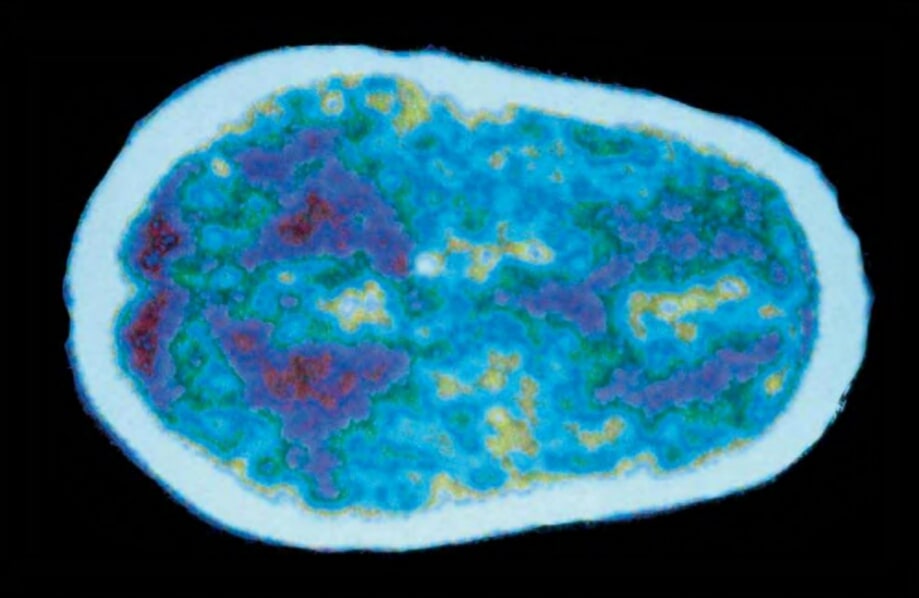


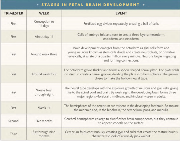
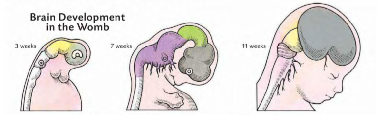
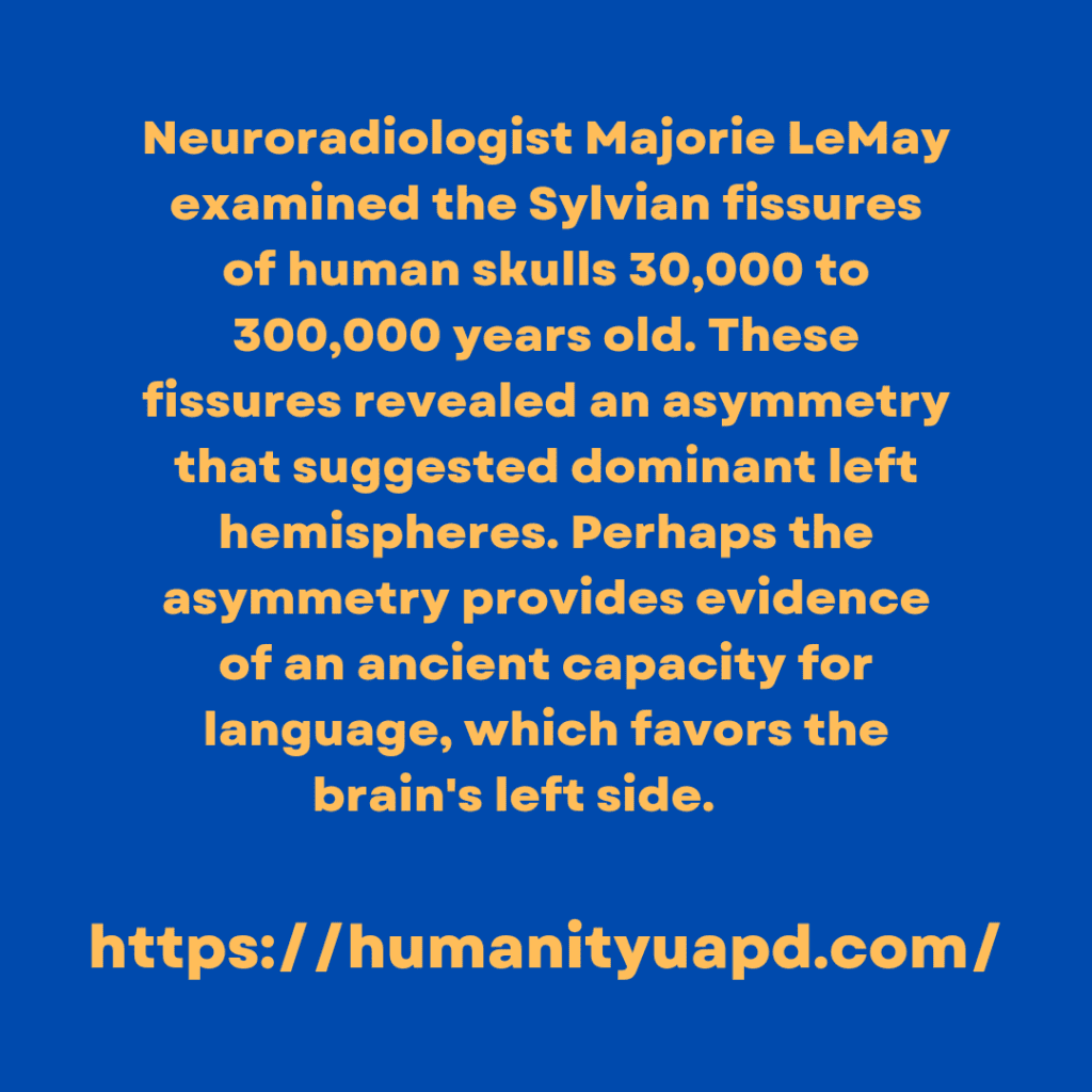
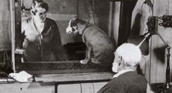
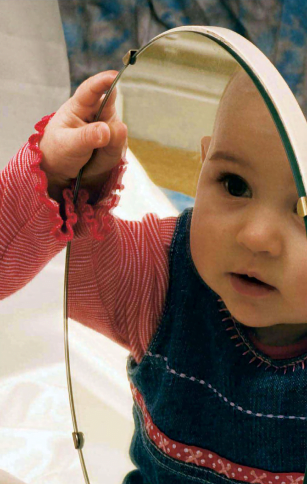
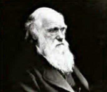
![DIABETES INSIPIDUS / DIEBETES MELLITUS [ CLASSIFICATIONS ] - Blueberries are rich in acetylcholine and antioxidants, making them an excellent food for brain health.](https://humanityuapd.com/wp-content/uploads/2022/11/IMG_20221112_163419.jpg)
![DIABETES INSIPIDUS / DIEBETES MELLITUS [ CLASSIFICATIONS ] - Regular tests help diabetics monitor levels of glucose in the bloodstream.](https://humanityuapd.com/wp-content/uploads/2022/11/Screenshot_2022-11-12-17-12-04-410_com.google.android.apps_.docs2_.png)

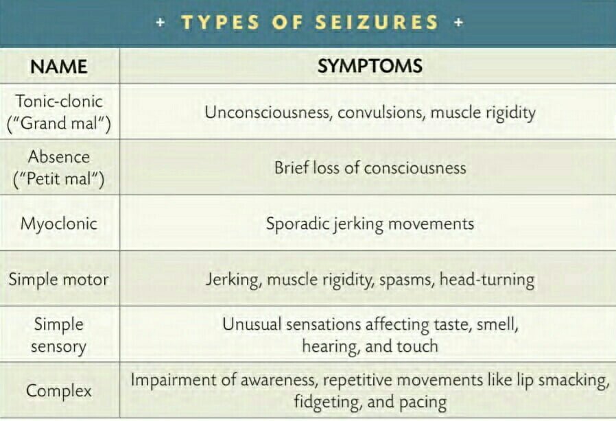

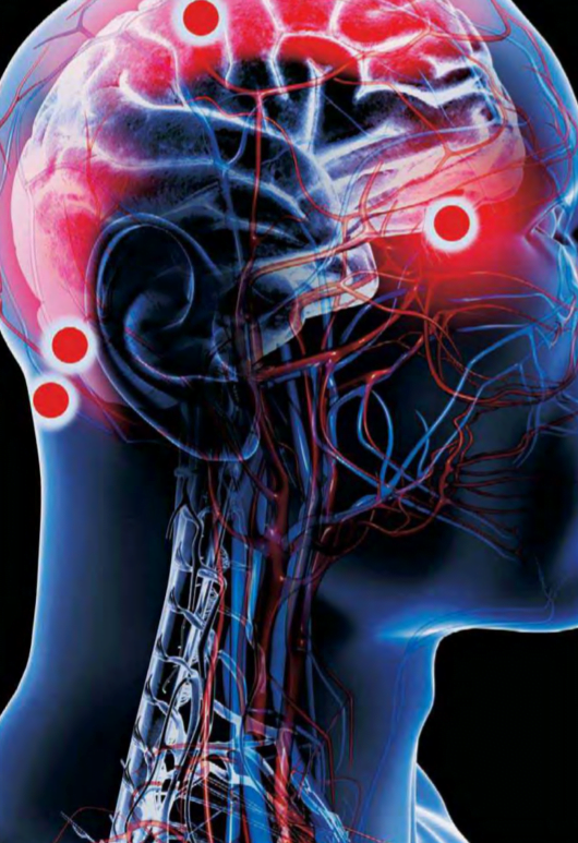
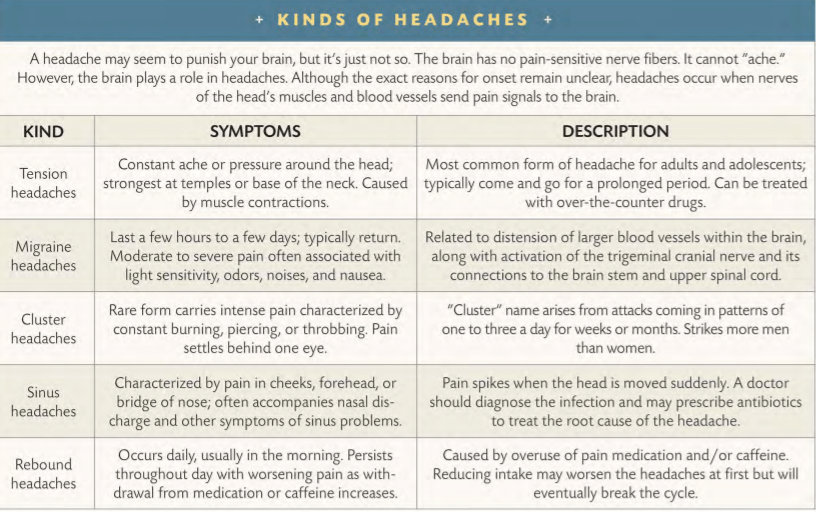
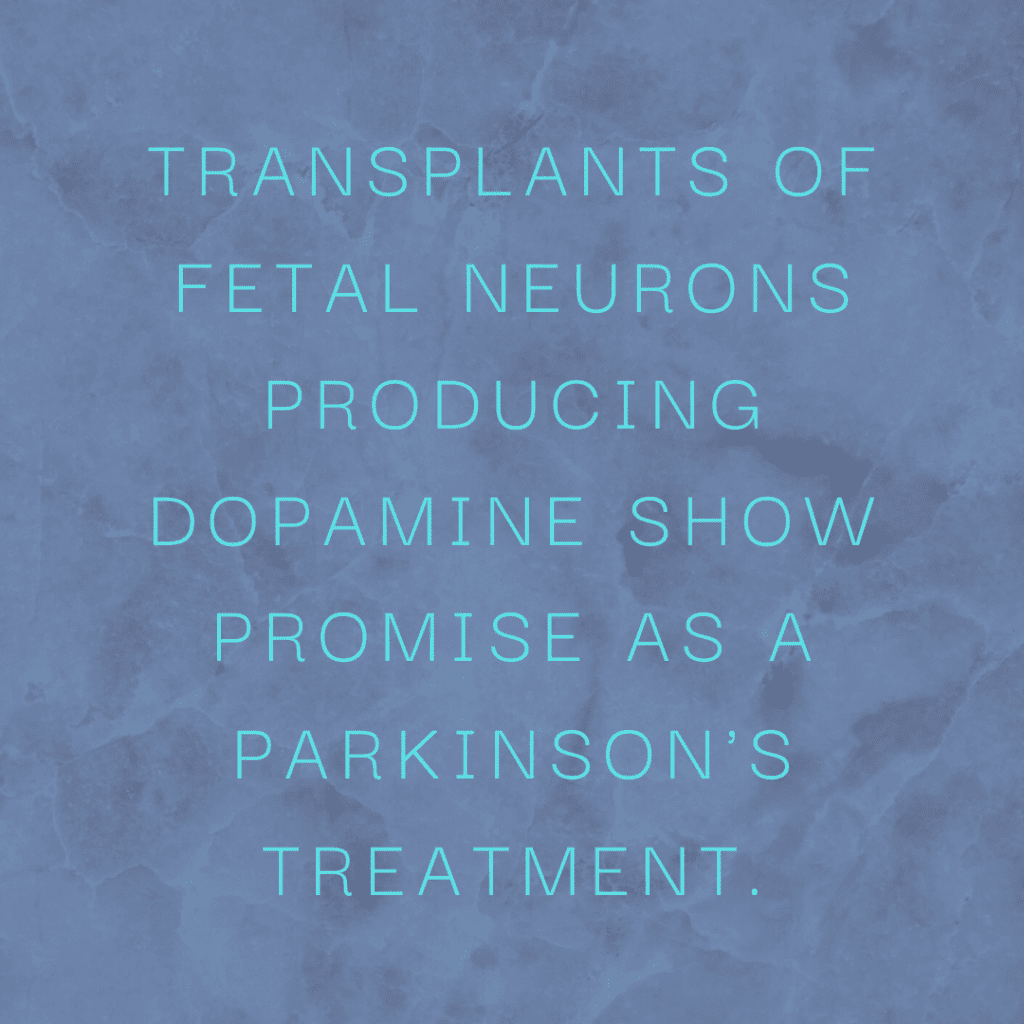
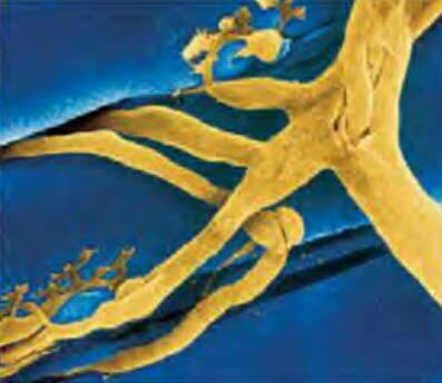

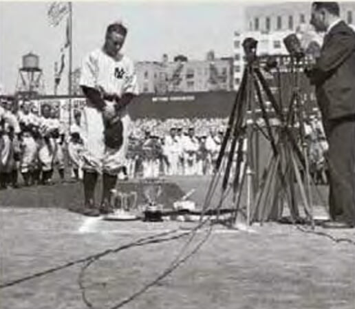


![GOOD FEELINGS / PLEASURE CENTERS [ THE NERVOUS SYSTEM ]](https://humanityuapd.com/wp-content/uploads/2022/10/Screenshot_2022-10-31-15-30-10-044_com.google.android.apps_.docs4_.png)

![PATHWAYS / GRAY MATTER [ MESSENGERS ( THE NERVOUS SYSTEM ) ]](https://humanityuapd.com/wp-content/uploads/2022/10/Screenshot_2022-10-29-15-14-24-029_com.google.android.apps_.docs_.jpg)
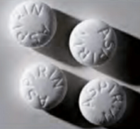
![PAIN GATEWAY [ MESSENGERS ( THE NERVOUS SYSTEM ) ]](https://humanityuapd.com/wp-content/uploads/2022/10/png_20221027_182415_0000-1024x1024.png)

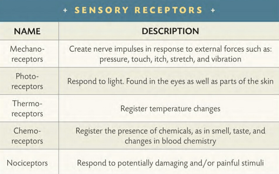
![THE NERVOUS SYSTEM [ HARMONY ]](https://humanityuapd.com/wp-content/uploads/2022/10/Screenshot_2022-10-23-20-28-12-530_com.google.android.apps_.docs_.jpg)
![THE AUTONOMIC NERVOUS SYSTEM / TWO BRANCHES [ HARMONY ]](https://humanityuapd.com/wp-content/uploads/2022/10/Screenshot_2022-10-22-16-53-42-536_com.google.android.apps_.docs_-472x1024.jpg)
![THE NERVOUS SYSTEM [ IN HARMONY ( MANY PARTS/HEAD & BODY ) ]](https://humanityuapd.com/wp-content/uploads/2022/10/Screenshot_2022-10-18-12-53-55-262_com.google.android.apps_.docs3_-525x1024.png)
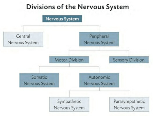

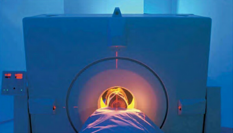
![BRAIN MAPPING [ LOOKING INSIDE ( THE AMAZING BRAIN ) ]](https://humanityuapd.com/wp-content/uploads/2022/10/IMG_20221014_204708.jpg)
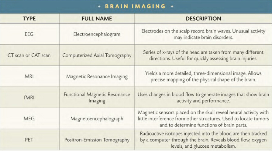


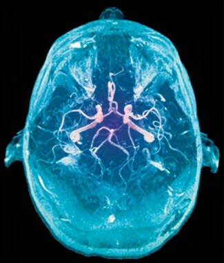
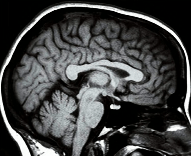
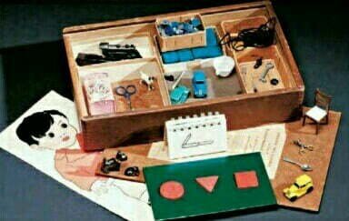
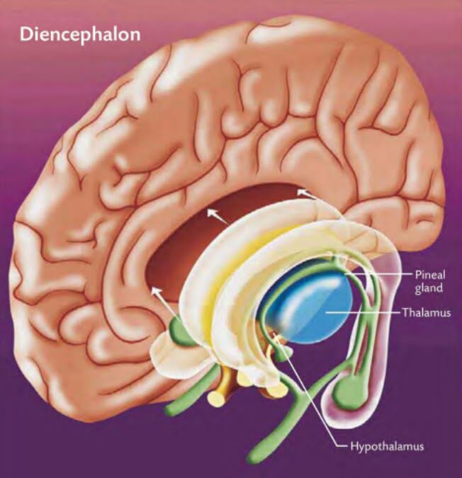
![ANATOMY [ DIFFERENT PARTS DIFFERENT RESPONSIBILITIES ( THE AMAZING BRAIN ) ]](https://humanityuapd.com/wp-content/uploads/2022/10/human-brain-vector-concept-vector-id10569719223.jpg)
![ANATOMY [ DIFFERENT PARTS & DIFFERENT RESPONSIBILITIES ( THE AMAZING BRAIN ) ]](https://humanityuapd.com/wp-content/uploads/2022/10/Screenshot_2022-10-07-12-59-20-065_com.google.android.apps_.docs_.jpg)
![NERVE CELLS - LIFE SPAN [ THE AMAZING BRAIN ]](https://humanityuapd.com/wp-content/uploads/2022/10/Screenshot_2022-10-04-13-05-57-139_com.google.android.apps_.docs4_.png)
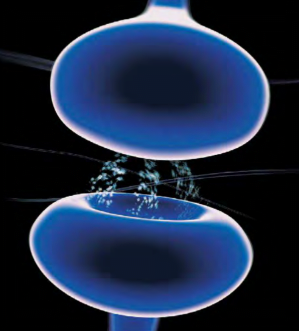
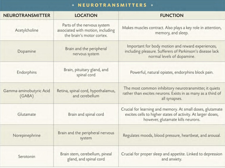
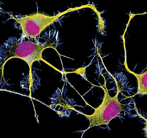
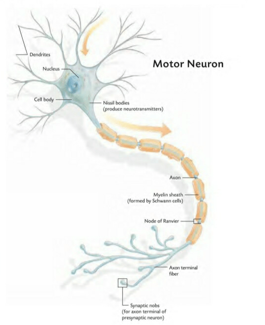


You must be logged in to post a comment.