Magnetic resonance imaging (MRI) gives a more detailed 3-D picture than a CT scan. An MRI relies on an intense magnetic field generated in a cylinder that surrounds the patient. It allows precise mapping of the physical shape of the brain. Its magnetic field is so powerful that it causes some of the atoms inside the brain to jerk into alignment. Then a series of radio waves from the MRI scanner bounce off the affected atoms and push them slightly out of line. When the energy from the radio signals is turned off, the atoms move back into their magnetic alignment, emitting telltale energy patterns along the way.
Addictive drugs work by mimicking neurotransmitters or altering their work. Brain scans reveal physical changes in the synaptic activity of a drug user. The drug known as Ecstasy, for example, can permanently damage neurons that produce serotonin.
Computers read these minuscule bits of energy and assemble images of cross-sections of the brain. Slices can be placed atop each other, like the layers of a cake, to represent the entire brain in three dimensions, or they can be examined individually, providing a closer look at localized phenomena. Comparisons of MRI scans of a single brain over time can show its growth-or reveal its deterioration.
WHEN THE DENTIST asked British philosopher Bertrand Russell where he felt pain, Russell replied, with humor and honesty, “In my mind, of course.” Russell knew the brain uses the senses to collect data about the world and construct a version of “reality.” Whether that world actually exists independent of the mind makes little difference to the sufferer of a tooth- ache-the pain hurts just the same. In fact, some philosophers, such as George Berkeley (1685-1753), have questioned whether “reality” exists.
In addition to mere structure, an MRI can also capture a snapshot of thought. A variation called a functional MRI, or f-MRI, builds upon the fact that a blood cell’s magnetic properties change according to how much oxygen it contains. Receptor cells use oxygen as they take in signals from surrounding cells; burning oxygen causes cells to require more oxygen-rich blood. As blood surges toward neurons where synapses are firing with thought, emotion, or other impulses, the oxygen they carry gives off a traceable signature of radio waves. Different thoughts light up different areas of the brain in an MRI. The processes of peaking, reading, appreciating humor and music, and recognizing faces illuminate various groups of neurons. MRI techniques thus help localize areas associated with certain brain functions.
WHAT IS INTELLIGENCE [ LOOKING INSIDE ( THE AMAGING BRAIN ) ]
PERHAPS NO scientific book of the past half century stirred up as much controversy as The Bell Curve: Intelligence and Class Structure in American Life. The 1994 book, by Richard ]. Herrnstein and Charles Murray, begins simply: “That the word intelligence describes something real and that it varies from person to person IS as Universal and ancient as any understanding about the state of being human.” From there, the authors delve into definitions of intelligence and how it can serve as a good predictor for success in life.
Then they argue that different levels of intelligence lead to social outcomes, instead of the other way around a person oflow intelligence is more likely to end up a criminal or unemployed, for instance and that intelligence levels have an observable correlation to biology.
Following the track linking genetics to intelligence, the authors make claims linking racial differences to intelligence, and thus the positive and negative social outcomes that define modern life. If a group of people can’t change their biology, goes this hypothesis, they cannot change their social outcomes.
Does the brain’s biology determine intelligence, and thus lock humans in to paths toward success or failure? It’s a potent question.
DEFINING INTELLIGENCE
Part of the problem lies in the definition of intelligence. Neuroscientists don’t agree on what the word means. Nor do they agree on what intelligence tests are actually measuring. Tests don’t measure motivation, persistence, social skills, and a host of other attributes of a life that’s well lived. Some say, only half facetiously, that IQ tests measure only one’s ability to perform well on IQ tests.
Studies of identical twins have shown that certain regions of the brain are highly inheritable, affecting overall intelligence.
Neurologist Richard Restak likes to deliberately cloud the issue during his lectures by showing students images of two PET scans. Each reveals the level of brain activity of a student doing a problem in a Raven’s Colored Progressive Matrices test, which aims to measure “fluid intelligence,” or the ability to solve an unfamiliar kind of problem. In one scan, the image is illuminated in red , and orange, representing an increase in brain activity. In the other, the cool shades of blue and green represent a less intense level of brain function. When Restak asks the students to guess which of the two students scored higher on the Raven’s test, and thus (one assumes) possesses superior intelligence, the students invariably pick the brain lighted up like a Christmas tree. Instead, the student with the less active PET scan posted a higher Raven’s score. The explanation: The brain that finds a problem easy to solve doesn’t have to work as hard.
TYPES OF SMARTS
There are several aspects of intelligence. Most are related, but historically not all have tested what they set out to test. For example, some early IQ tests measured knowledge of facts, which actually is a function of education and memory rather than the ability to reason. In general, however, a person’s performance on a test of fluid intelligence is a good predictor of performance on a wide range of mental exercises. For example, increased fluid intelligence correlates to a high level of “working memory”-one’s ability to remember information temporarily which can range from remembering where you parked your car to which words or number combinations you tried and rejected in doing a crossword puzzle or Sudoku. People with powerful working memories are more focused in solving problems.
Scientists use the term “g-factor” when discussing the general measure of mental ability, found in vocabulary size, mechanical reasoning, and arithmetical computations. They relate it to the properties of efficient neural functioning, rather than the value of knowledge in its own right. The prefrontal cortex, right behind the forehead, is the most likely home for much of the neural processes associated with one’s g-factor abilities. When it’s damaged, a person suffers a variety of impairments to abstract reasoning, and it lights up during brain scans taken during a variety of intelligence tests.
“You have less frontal development than I should have expected,” says the evil Professor James Moriarty when he first lays eyes on Sherlock Holmes in a story by Arthur Conan Doyle. As scientists have discovered, the size of the prefrontal cortex in healthy brains generally correlates to fluid intelligence. (Perhaps Moriarty subscribed to the theory of phrenology and believed cortex size correlated to the bulging of a forehead. It’s not so.)
Psychologist John Raven devised the Raven’s Colored Progressive Matrices Test in 1938, a non-verbal test of intelligence in children.
But the size of a cortex doesn’t mean, QED, that biology causes intelligence the same way gravity causes an apple to fall. Identical twins vary in their performance on IQ tests. In some cases, one twin develops schizophrenia or some other disorder, and the other does not. Furthermore, when identical twins are separated at birth and raised separately in similar environments, they show only a 72 percent correlation in intelligence.
FAMILY INFLUENCE
At best, genetics accounts for only a substantial fraction of intelligence. Perhaps heredity sets an upper limit for intelligence (through the potential ability to make neuronal connections), which then becomes subject to other forces. An environment with plenty of books and challenging toys plays a key role in increasing aspects of a child’s intelligence but so does willingness to exercise the brain. Political scientist James R. Flynn noted that IQ scores have dramatically increased over the past several decades in many countries. He attributes the so-called Flynn effect to increases in modern humans’ greater ability to solve abstract problems, possibly from living in a more intellectually stimulating world.
The brain’s ability to rewire neuronal networks no matter how old the nerve cells provides the means to improve mental function. Instead of looking at family or ancestral heritage and deciding it determines mental performance, humans can set about learning new skills and tasks. Challenging the brain may not raise the score on a particular IQ test, but it will help the brain to perform better.
Scientists have long Dreamed of Exammmg how the brain works within a living body. The problem, though, was figuring out how to get inside the head without causing injury or even death. Doctors treating wounds from wars and accidents have been able to get glimpses of living brain tissue, but aside from poking or prodding, have had little to do with experimental observation.
Some early noninvasive attempts included phrenology, the pseudoscience developed in the early 19th century that measured the bumps on the outside of the skull as a means of analyzing the mental powers and characteristics. They stemmed from the theories of a German doctor, Franz Joseph Gall, who argued in the late 18th century that the separate faculties of the brain must manifest themselves in the shape of the overlying bone. Phrenology’s popularity peaked between the 1820s and the 1840s but soon waned as the century progressed.
Overall, at least half of all cases of dementia-formerly known as senility can be traced to Alzheimer’s disease.
Toward the end of the 19th century, a new method of probing the hidden workings of the brain arose, again in central Europe. Wilhelm Wundt, known as the founder of experimental psychology, created a laboratory in the mid-1870s in Leipzig to perform research into psychology. The word derives from the Greek psyche, meaning “mind” or “soul.” Wundt considered his research a way to get at the workings of the mind, which many still considered to be separate from the tissue of the brain.
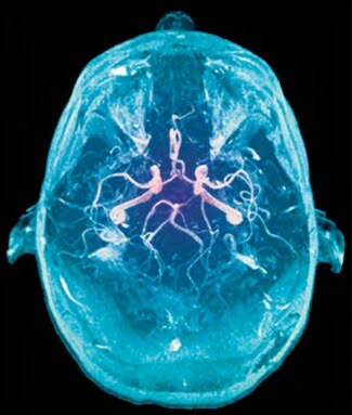
An angio-MRI of a 27-year-old woman reveals the arteries that provide oxygen to her brain.
In particular, Wundt aimed to examine the elements that made up consciousness and explain how they worked together to create the mind. Wundt concentrated on stimulus-response experiments, as he considered sensation the contact point between the external, physical world and the inner, psychological world. He recorded when and how sensations entered consciousness, including such mundane facts as whether one musical tone sounded higher or lower than another one did.
A contemporary of Wundt’s, the American William James, also took up psychology as a tool to probe the mind. India his famous 1890 textbook The Principles of Psychology, James described processes including the sense of self, memory, movement, and sensation.
Your brain uses about 12 watts of ” power-a fraction of the energy of a household lightbulb.
Assessing the brain’s performance through intelligence testing was another way science attempted to access the living brain. In the 1900s, French psychologist Alfred Binet created the first IQ test as a way to measure intelligence. That test, designed to see which French schoolchildren needed special assistance, became the genesis of all IQ tests that followed.
Meanwhile, in Austria, Sigmund Freud (1856-1939), the founder of the psychoanalytic school of psychology, turned his interest in neurology into the study of the workings of the brain and the ways in which they affect behavior. He predicted, correctly, that someday the study of the physical workings of the brain would dovetail with his observations about unconscious drives.
LOOKING INSIDE [ SEEING THE BRAIN AT WORK ( THE AMAGING BRAIN ) ]
ONCE THE brain’s true purpose was ascertained, scientists began finding new ways to observe it and its functions. Starting with noninvasive methods, like IQ tests, they tried to learn more about the living brain and measure how it worked. These intelligence tests painted a picture of how the brain collected information, processed it, and then made conclusions.
CT scans open windows into the brain’s interior structure.
Peering inside a living brain was virtually impossible-most of what scientists knew abour the brain’s anatomy was based on autopsies. But in the late 19th century, the invention of the x-ray made it possible to take a look inside the skull. In the 20th century, new scanning methods came along and gave greater insight into how the living brain works.
TESTING INTELLIGENCE
ALFRED BINET (1857-1911) made the first serious effort to chart intelligence. In 1905, France commissioned him to create a test to identify students whose intelligence was below average. Binet and his doctoral student, Theodore Simon, devised a series of tasks for children. They then tested how well children of various ages performed the tasks, which gradually increased in complexity. Their work led them to create a scale of normal mental functioning. Binet’s intelligence scores compared a child’s mental abilities with those of h is or her peer group. The test has been updated many times.
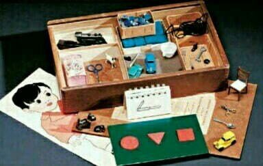
A 1937 Stanford-Binet intelligence test includes miniatures and printed matter.
During World War II, the American government gave Army recruits intelligence tests to screen them for war work. Plenty of other groups have been given IQ tests since then, allover the world. If you look only at their scores, you might think humans are getting smarter all the time. New Zealand political scientist James R. Flynn observed that standardized intelligence test scores from 20 countries historically have kept rising by about three points a decade. The reason isn’t entirely clear, but it’s possible that improvements in nutrition, coupled with the more stimulating environments in which children are raised, contribute to greater neuronal complexity.
Today, scientists still wrestle not only with what intelligence is, but also how it can be measured. Harvard University’s Howard Gardner believes at least seven types of intelligence exist, from the mathematical to the athletic.
ANATOMY [ DIFFERENT PARTS & DIFFERENT RESPONSIBILITIES ( THE AMAZING BRAIN ) ]
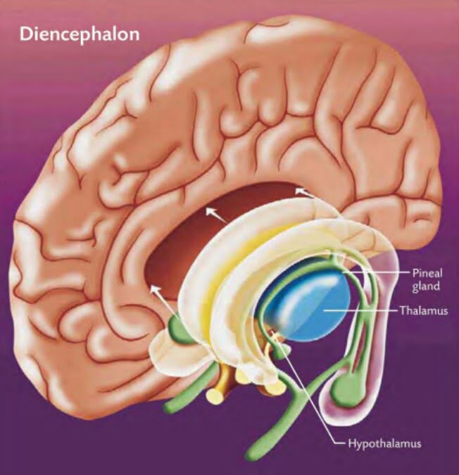
DIENCEPHALON
In the center of the brain, between the cerebrum’s two hemispheres, lies the diencephalon. It consists largely of three important structures : the Thalamus, Hypothalamus, and Epithalamus. The Thalamus acts as a relay for sensory information on its way to the cerebrum and is crucial to memory and emotions. The tiny Hypothalamus exerts control over the autonomic nervous system and performs other functions, including regulating body temperature.The Epithalamus includes the pineal gland, which drew Descartes’s attentions. Instead of housing the soul, scientists now know it helps to regulate the body’s rhythms of sleeping and wakefulness.
Elements of the diencephalon link THE you left and right hemispheres.
CEREBELLUM
At the back and bottom of the skull rests the cerebellum. Like the cerebrum, it too is divided into halves and deeply fissured. Its role is to coordinate movement and balance. Precise physical activities that must be practiced to be performed well-hitting a golf ball, doing gymnastics, picking a pattern of notes on the strings of a guitar-are processed in the cerebellum. The cerebellum also is known to play a role in emotion and action.
Misunderstanding of the work of neuroscientist Roger Sperry in the 1970s fed the notion that everyone is either “left brained” or “right brained.” Although each hemisphere has special functions, the two halves work closely together in a healthy mind. Humans are whole brained.
MEDULLA OBLONGATA
Where the brain meets the spinal cord is the brain stem. The spinal cord, the central route of nerve cells connecting brain and body, terminates in a 1.2 inch extension into the lower brain known as the medulla oblongata, home to motor and sensory nerves. Here is where the nerves from the body’s left and right sides cross each other on their way toward the cerebrum. Basic body functions such as heartbeat and respiration are controlled in the medulla.
Above the medulla lie the pons and midbrain. Pons means “bridge,” and that’s what it does-it acts as a bridge between the medulla and other brain regions. The midbrain links the pons to the diencephalon and controls reflexes of the ear and eye, such as the jolt the body experiences when startled.
FUELING THE BRAIN
Blood pumped from the heart pushes upward into the brain through two main sets of blood vessels, the internal carotid and vertebral arteries. Spiderwebs of smaller vessels, like distributary waterways at a river’s mouth, send blood into every region of the brain.
The brain uses oxygen III a hurry. While the brain weighs only about three pounds, a mere fraction of body weight, it burns 20 percent of the body’s oxygen and glucose. Most of that energy is mere upkeep, keeping the brain on the razor-sharp edge of action by maintaining the electric fields of the membranes surrounding the synaptic clefts. Actually thinking adds very little to the demand for energy-a fact that is somewhat counterintuitive for anyone who has ever struggled with a particularly difficult math problem or foreign language translation.
To get fuel to hungry brain cells, the body relies on the constant circulation of glucose. It’s a kind of sugar that circulates via the bloodstream. Neurons can’t stock-pile glucose like coins in a bank, so they require a ready supply of this source of chemical energy. Neurons use the fuel of glucose to manufacture and transport molecules of neurotransmitters and enzymes. They also use plenty of energy- half of the brain’s total, in fact-to transmit electro-chemical signals from cell to cell. The body obtains glucose from starches and sugars in the daily diet. Good sources include grain, fruits, and vegetables. During periods of intense concentration, glucose levels decline in brain regions associated with memory and learning. Such a decline can cause a feeling of fatigue in the body and the brain.
AN OLD BRAIN can be an amazingly healthy and creative one. Consider:
Ben Franklin left public service at age 82.
Mary Baker Eddy founded The Christian Science Monitor at age 86.
Robert Frost published his last collection of poems at age 88.
George Bernard Shaw was still writing plays at age 94.
Grandma Moses received a painting commission at age 99.
ANATOMY [ DIFFERENT PARTS DIFFERENT RESPONSIBILITIES ( THE AMAZING BRAIN ) ]
THE FRONTAL LOBE
A portion of the frontal lobe of each hemisphere called the precentral gyrus controls the body’s movements. Oddly, each hemisphere moves the opposite side of the body, as if the brain’s wiring some-how became crossed. Hence, the movements of the right hand and right foot, as well as the rightward gaze of both eyes, are governed by the left side of the brain. This phenomenon has been observed for centuries. Hippocrates noted that a sword injury to one side of the head impaired movement on the body’s opposite side. And while observing combat wounds during the Prusso-Danish War of 1864, German doctor Gustav Theodor Fritsch noted that if he touched the cerebral cortex as he dressed a head wound, the patient twitched on the opposite side of his body. If one hemisphere’s precentral gyrus is destroyed-during a stroke, for instance-paralysis will result in half the body.
In front of the precentral gyrus lie the premotor cortex and the prefrontal fibers. The former organizes the body’s complex physical movements, whereas the latter inhibit actions. Inhibition is useful in a variety of social settings, such as preventing shouting in a quiet movie theater.
THE BRAIN NEEDS regular exercise if its neurons area to remain sharp. Repetition of newly learned tasks helps make those new connections stronger. Without stimulation, dendrites recede and the brain settles into simpler patterns of operation. Neurologist Robert Friedland has shown that posing new challenges to the brain can help in the defense against Alzheimer’s disease.
Perhaps not surprisingly, “Use it or lose it” appears TO be true not on Iy of mental exercise but also of physical stimulation of the brain. The brain is like other organs and works better when the body is healthy. Exercising the body regularly appears to help ward off Alzheimer’s disease, as do reducing body weight, lowering blood pressure, and eating a more healthful diet. General exercise that builds up cardiovascular endurance improves blood flow to the brain. A healthy heart usually is linked to a healthy brain, especially in the brain’s “executive function, ” which is crucial to a slew of mental tasks.
A combination of physical exercise and mental gymnastics protects the brain against deterioration with age. To spur on the brain to make new neuronal connections and protect the ones it has, there are a number of activities to try, such as: ~ Learning a new language . ~ Listening to classical music. ~ Solving mental puzzles and games, like crossword puzzles and Sudoku . ~ Eating a healthful diet. ~ Walking, jogging, or cycling regularly to promote cardiovascular health . ~ Maintaining a healthy weight.
PARIETAL LOBE AND TEMPORAL LOBE
In the parietal lobe lies the somatosensory cortex, which takes in stimulations of touch and other sensations. While lower parts of the brain register pain and pressure, the sensory cortex helps localize such feelings. Damage to the sensory cortex may result in confusion about which part of the body may be registering pain.
The temporal lobe is home to the functions of hearing and appreciation of music and to some aspects of memory. Self-experience also resides in this lobe. Electrical stimulation of the temporal lobe may dredge up intense feelings from the memory-the experience of reliving the past, known as deja vu-or do just the opposite, causing familiar people and objects to become unrecognizable.
At its base, the temporal lobe connects with the limbic system, a series of brain structures also known as the animal brain. This system allows humans to experience intense emotions such as anger and fear as well as react to these feelings.
OCCIPITAL LOBE
Behind the temporal lobe, near the rear of the head, lies the brain’s visual center in the occipital lobe. Far from the eyeballs, which takes in visual information, this portion of the cerebral cortex processes electrical impulses that begin with light waves striking the retina. Wounds to the back of the head injuring the visual cortex can sometImes cause blindness.
ANATOMY [ DIFFERENT PARTS & DIFFERENT RESPONSIBILITIES ( THE AMAZING BRAIN ) ]
FOUR DIVISIONS
Moving inward, we come to the organ itself. The brain may appear to be a Ulllform mass of folded, pink tissue. But a closer look reveals different lobes, regions, structures, and parts that all help regulate body functions, interpret information from the body, and react to stimuli. The brain has four main parts: the cerebrum, diencephalon, cerebellum, and brain stem.
SHAKESPEARE WEIGHS IN on the human brain in his plays:
“Tell me where is fancy bred, Or in the heart, or in t he head?”-The Me rchant of Venice
“The brain may devise laws for the blood, but a hot temper leaps o’er a cold decree.” – The Merchant of Venice
“Her beauty and her brain go not together. ” – Cymbeline
“He has not so much brain as ear-wax.” – Trai/us and Cressida
CEREBRUM
This largest, topmost layer of the brain is the cerebrum. It’s what most people visualize when they use their brains to picture their brains. The external layer is called the cerebral cortex. Its outer por- tion is gray from the presence of billions of nerve cell bodies, while the inner portion is white from the tangle ofaxons coated in their myelin sheaths.
In 1999, scientists discovered that Albert Einstein’s inferior parietal lobe, associated with mathematical and spatial reasoning, was 15 percent wider than that of an average brain.
In the cerebral cortex lies the core of information processing that separates humans from other animals, including reason, language, and creative thought. Homo sapiens has more of its brain in the cerebral cortex-approximately 76 percent-than any other animal. (Chimpanzees rank second at 72 percent, while dolphins have only 60 percent.)
FISSURES AND HEMISPHERES
The cerebrum is divided into parts by deep fissures. The largest of the brain’s fissures is immediately evident to the naked eye. Down the center of the cerebrum, separating it into left and right hemispheres, is the longitudinal fissure. The left and right halves of the cerebrum appear to be nearly mirror images of each other.
While they look alike, the two halves perform and control very different functions. The left hemisphere long has been considered the dominant half because of its role in processing language, but the right hemisphere is gaining new attention for its role in emotions and spatial cognition, as well as the integrative function that helps bring bits of information together to create a rich image of the world.
Connecting the two hemispheres are bands of nerve fibers that allow information to be passed back and forth between the two halves of the brain. The largest bundle, containing about 200 million nerve fibers, is the corpus callosum.
Two divides known as the Sylvi an fissure and central sulcus lie on the outside edges of the hemispheres. Their locations serve as boundaries on a map, dividing the hemispheres further into four lobes. The frontal lobe lies forward of the central fissure. Between the Sylvian and central fissures are two lobes that merge together, the parietal followed by the occipital. Behind the Sylvian fissure is the temporal lobe.
ANATOMY[ DIFFERENT PARTS, DIFFERENT RESPONSIBILITIES( THE AMAZING BRAIN ) ]
THE FIRST STEP to a better understanding of the brain is getting acquainted with its parts. From the protective structures on the outside to the hardworking parts on the inside- knowing where each structure is and how it interacts with the world gives greater insight into brain function and the problems that mayanse.
The eight bones that form the cranium shield the brain from injury.
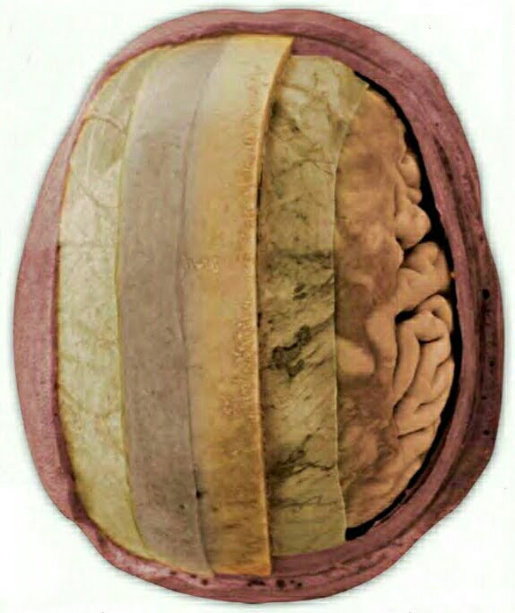
PROTECTION
To take a tour of the human brain, begin with the crown of the skull, a collection of 22 bones that house the brain and protect it from harm. Except for the mandible (or jawbone), all of these bones are fused together and immovable. The topmost and rearmost bony parts form the cranium, the brain’s tough, protective shell.
Inside, three membranes present themselves to provide more layers of protection. Immediately under- neath the skull is the dura mater, Latin for “hard mother.” The next layer, the arachnoid, overlays the brain’s network of crevasses. Early observers likened it to the spun lace of a spider, giving it a name that means “cob- web.” The lowest of the three membranes, the pia mater (“tender mother”), is filled with tiny blood vessels. It embraces the brain surface like a mother cradling a child in her arms; every dip and rise in the brain matter is form-fitted by the pia. The ridges are called gyri, which means “twisters,” while its grooves are sulci, or furrows.
BRAIN CUSHION
Flowing between the arachnoid and pia membranes IS the brain’s cerebrospinal fluid. This liquid bathes the brain’s gyri and sulci, including the deepest grooves, which are known as fissures. Fluid- filled ventricles-the hollows that some philosophers such as Thomas Aquinas considered home to the mind-curve deep into the brain and connect to the spinal cord’s central canal. Cerebrospinal fluid cushions the brain, provides nourishment for tissues, and perhaps acts as an internal channel of chemical communication.
Layers of coverings combine to cushion, protect, and support the brain.
Poet Lord Byron’s brain weighed 79 ounces, well above the average human brain’s weight of 48 ounces.
PROTECTION
The body has evolved formidable defenses to protect its most vital organ. While capillaries in other parts of the body allow cells to absorb harmful substances from the blood, the brain has the so- called blood-brain barrier with only limited permeability. Thick, tight membranes in the brain’s blood vessels screen out many substances in the bloodstream. Crucial chemical such as oxygen and glucose can cross into the brain, as well as a few harmful ones, such as alcohol and nicotine. Frustratingly, many beneficial chemical compounds, such as drugs designed to attack tumors, are turned back.
Amazingly, the cells that perform the complicated ballet of electrochemical transmission can live more than a hundred years, but they do not get replaced like most other body cells. Except for the hippocampus and the olfactory bulb, where new neurons have been shown to grow from stem cells, the neurons a person has at birth are all he or she will ever have. During the busiest times of neuron generation in the developing brain of a Fetus, about a quarter million neurons are created every minute. They start from precursor cells and then migrate and differentiate.
When a neuron in the central nervous system dies or its long fibers are cut, it does not regen- erate. Medical science currently has no cure for catastrophic nerve injuries of the spinal cord, and once a major communication line to or from the brain has been cut, it cannot be repaired. But new research with neural stem cells sug- gests neurons may yet be coaxed into regeneration.
REEVE’S RESEARCH
RESEARCH INTO HOW TO regenerate nerve tissue after injuries like transections, a complete severing of the spinal cord, owes a great deal to the late actor Christopher Reeve. In 1995, Reeve shattered a cervical vertebra in a horseback riding accident and became paralyzed from the neck down, a condition known as quadriplegia. The injury was not quite a transection-he eventually regained some sensation-but nevertheless proved devastating. His public appearances in a wheelchair until his 2004 death drew attention to spinal injuries and ultimately raised millions of dollars to help seek a cure for nerve damage.
Tim Berners-Lee, a creator of the World Wide Web, likens the brain’s complexity to the nearly infinite capacity for Web sites to connect to each other. “A piece of information is really defined only by what it’s related to,” he said. “The structure is everything. There are billions of neurons in our brains, but what are neurons? Just cells. The brain has no knowledge until connections are made between neurons. All that we know, all that we are, comes from the way our neurons are connected.”
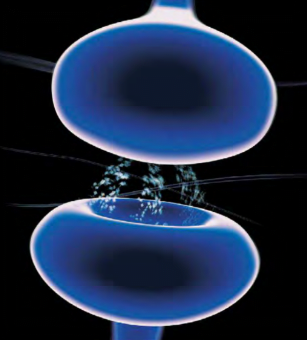
Communicating with another cell, neurotransmitters journey across a synapse.
Transmissions between neurons take place in two stages. The first is electrical. An electrical discharge travels the length of an axon. When it reaches the axon terminal that abuts the synaptic space, it sets the second stage in motion. This but- ton, like the rest of the nerve cell, has an outer wall called a mem-brane. Its envelope contains a solu- tion of messenger chemicals. These electrically charged chemicals move in the solution, constantly poised to respond to an impulse and exit through small openings of the membrane and into the synapse. When an electrical impulse arrives from the axon, if it is of sufficient strength it trips a trigger that releases one of the messenger chemicals, called a neurotransmitter, from storage in the button.
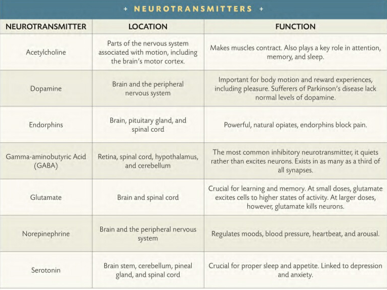
NEUROTRANSMITTERS
The neurotransmitting chemical then enters the synapse. Like a ferryboat crossing a small stream, the neurotransmitter traverses the synaptic cleft and attempts to link up with the dendritic membrane of a receptor cell. The journey across the synapse takes only a thousandth of a second. The receptor cell’s surface contains specially shaped docking sites, so particular neurotransmit- ters can dock only at the appropri- ate places, just as a key needs exactly the right shape to fit into a lock. The neurotransmitter either excites the receptor cell into action or dampens it into inaction. Once the receptor cell has been stimulated by the neurotransmitting chemical, the communication reverts to an elec- trical signal. It travels the length of the new cell until it reaches the synapse of another receptor cell, and starts the process all over again. After they have done their job in the synaptic space between nerve cells, neurotransmitting chemicals are reabsorbed by the transmitting neuron and prepared for rerelease (a process known as reuptake) or broken down and metabolized by enzymes in the synaptic space. It sounds like a lot of work, but neurons can repeat the electrochemical firing process up to a thousand times a second.
WAKING IN THE middle of the night on the eve of Easter, 1921, German-born pharmacologist Otto Loewi (1873-1961) recalled an inspiring dream that gave him an idea for an experiment that would shatter scientists’ conception of neural communication.
Most turn-of-the-century brain Scientists believed nerves sent impulses via electric waves, firing sparks across the synaptic gap, neuron to neuron. In this way, they thought, motor intentions born in the cerebral cortex could be transmitted to receptor muscles and organs throughout the body. Only a handful of scientists-most notably Loewi and his English counterpart, Henry Daleargued that chemical neurotransmitters are released at the synapse. An accelerant, noradrenaline, causes the heart to beat more quickly, Dale said. An inhibitor, acetylcholine, induces the opposite. Yet Dale was unable to extract either chemical organically, and lacking proof, his case remained dormant.
Then, as Loewi recalled, a fateful frog experiment flashed to him in a dream, and he dashed to his laboratory. He began with two frogs’ hearts. Stimulat- ing the vagus nerve of one to slow its beating, he applied a residual solution from this donor to a second heart, from which he’d severed the vagus nerve. The second heart immediately slowed, as if discouraged by an unseen force. Loewi’s hypothesis was correct: A neurotrans- mitter (acetylcholine) had slowed the first heart, leaving a trace fluid-enough to slow the second, isolated heart.
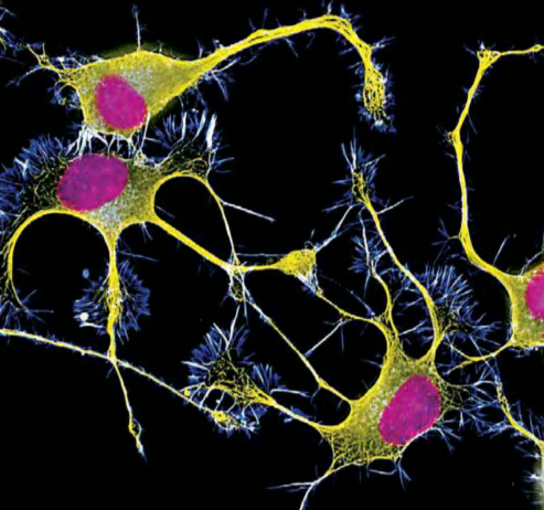
Precursors to axons and dendrites, in yellow and blue, respond to nerve growth stimulation.
The brain devotes huge amounts of neural circuitry to the hands, lips, and tongue.
Dozens of neurotransmitters have been identified, and more discoveries are expected. Certain neurotransmitters make muscles contract, help regulate sleep, and block pain. Research into the role of neurotransmitters in mental and physical health is constantly expanding, and neurotransmitter disorders have been linked to Parkinson’s disease, depression, Alzheimer’s disease, schizophre- nia, and a host of other illnesses.
NEURONS AT WORK [ CONNECTIONS / ACTION / GROWTH & SUPPORT / PLASTICITY ]
Neurons serve different functions. Motor neurons carry impulses to activate glands and muscles. Sensory neurons send impulses from the skin and other body parts to the central nervous system. Interneurons, residing in the brain and spinal cord, integrate the signals and are crucial in making decisions. Thus, neurons allow for information from the body to reach the brain, be processed, and sometimes result in responses.
Some liken the neuron to an old- fashioned, landline telephone. The body of the neuron compares to the body of the phone, where sig- nals are processed. The telephone receiver compares to the dendrites and their ability to gather informa- tion. And the axon compares to a telephone line, sending informa- tion processed in the phone body along an electrically conductive wire. It has the potential to pass information along to any other phone on the planet
NEW CIRCUITS
IF NEURONAL CIRCUITRY rewires itself in response to stimulation, do the brains of teens raised on the Internet and high-tech gadgets differ from those of older genera- tions? The answer most likely is yes. UCLA psychiatrist Gary Small believes tech-savvy children strengthen synaptic connections for electronic communica- tion while their circuitry for a face-to-face world, such as reading body language, fades. Meanwhile, late adopters of technology lag in their ability to master new communication media.
MAKING CONNECTIONS
The human brain contains ill the neighborhood of 100 billion neurons. Each neuron reaches out toward others with an array of dendrites and axon terminals. Each is capable of communicating with any other and, in the process, forging thousands of synaptic connections through the thickets of dendrites and axon terminals. All told, the brain has hundreds of trillions of synapses. No computer can match the human brain for its complexity and its potential for creative thought.
Communication occurs where two neurons come together. Camillo Golgi, a contemporary of Ramon y Cajal’s, believed that neurons physically touched each other, forming a continuous net- work of neural fibers. Ramon y Cajal disagreed. In his sketches, he painstakingly drew neurons whose dendrites invariably terminated at a tiny gap that prevented them from touching other neurons. His drawings did not lie.
In the synaptic cleft, a neuron communicates with its neighbors by issuing electrochemical commands that may be strictly localized or extend the length of the longest chains ofaxons.
PLASTICITY
Neurons are not physically bound to each other like so many lengths of pipe, so they have the flexibility to make, break, and remake relationships with other neurons. The ability to reshape neural interac- tions in the brain is referred to as plasticity. The brain’s ability to rewire itself helps it stay sharp.
The number of synapses may be as high as one thousand trillion, or the number 1 followed by 15 zeroes.
As the brain ages, it loses individual neurons, but it retains its power to form new connections that increase the mind’s complexity. In short, if new educational experiences challenge the brain to form new synaptic connections, its neurons will do more with less.
Experimental data with labora- tory animals demonstrate the principle of “use it or lose it.” When lab animals are placed in an environ- ment with challenging toys, their brains develop a far greater number of neuronal connections than those raised in a dull environment. The brains of animals from stimulating environments will even weigh more because of the greater number of synapses.
ANATOMY OF A NEURON [ NERVE CELLS ( THE AMAZING BRAIN ) ]
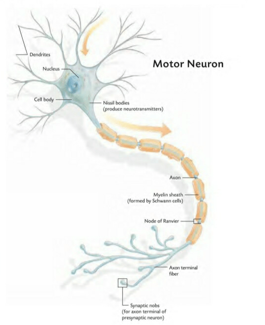
Each neuron has a main cell body. Like all cells, the neuron contains a nucleus and an exterior mem- brane, which sometimes receives electrochemical messages from other neurons. Chains of neurons send messages from the body to the brain: “Here is pain, in the left wrist.” “Here is the odor of soup.” “Here is a stony surface beneath the feet.” Chains also send mes- sages from brain to body: “Shake your hand “”Eat. “”‘Take a step. “
Each neuron has an array of branching fibers called dendrites that extend outward toward other neurons. Dendrites expand the surface area of the neuron, increas- ing its sensitivity to its neighboring neurons. While some neurons have only a few dendrites, others have hundreds. They act as receptors for signals traveling from other neurons, carrying information toward the main body of the nerve cell.
Each neuron also contains one electrically sensitive fiber called an axon extending from one end of the cell body. Axons may be as short as a fraction of an inch or as long as several feet, as is the case with axons extending from the spine to the toes. At the axon’s terminal end, as many as 10,000 branches spread out toward the dendrites of other neurons. Every branch terminates in a knoblike projection, like the business end of a paper match. These bulbs are called axon terminals, synaptic knobs, and boutons, or buttons.
Santiago Ramon y ajal, in a 1906 portrait, documented the existence of synapses.
OFTEN THE spirited competition between two great minds can yield amazing discoveries. Such was the case between Spanish neuroscientist Santiago Ramon y Cajal (1852-1934) and his Italian contemporary, Camillo Golgi (1843-1926), who shared the Nobel Prize in physi- ology or medicine in 1906. Ramon y Cajal was recognized for his deduction on the anatomy of a neuron; Golgi, for the staining process that made that deduction possible. Like most scientists at the time, Golgi held that neurons operate as one continuous, tangled network. Nerve cells must be fused, he said, to pass electrical impulses. Ramon y Cajal, howeveG envisioned chemical codes traveling across a synaptic gap between a single axon and the dendrites of the next cell. In 1887, Ramon y Cajal learned of Golgi’s staining technique and realized its superiority. He modified it, finding it worked well with thicker sections of nervous tissue. Bird sam- ples and tissue from younger animals were best, he surmised, because their axons lacked the protein sheath that obscures most nerve fibers. When impregnated with silver nitrate and viewed by microscope, these nerve cells jumped out as inky strokes on a yellowish background. La reazione nera-“the black reaction,” as Golgi called it-illuminated the infinitesimal as well as the road toward Ramon y Cajal’s revelation.
Around the length of most axons lies a special wrapping of fatty tissue called a myelin sheath. The sheath is formed by two kinds of glial cells, called Schwann cells in the peripheral nervous system and oligodendrocytes in the central nervous system. The wrap is not continuous; small gaps called nodes of Ranvier separate the cylinders of fatty tissue that surround the axons. The axon’s encompassing myelin acts as insulation, speeding the transmission of information in the form of nerve impulses moving at 9 to 400 feet per second.
When an electrical impulse reaches an axon terminal, it communicates across a tiny gap, called a synapse, separating it from the dendrite of another neuron. A few can connect directly with tissues of the skeletal muscles and glands, allowing direct communication.
Neurons differ in shape and complexity. Most, in particular the vast majority of those in the brain, are multipolar-they have one axon and a multitude of dendrites. The rest of the neurons are bipolar or unipolar. The former can be found in the retina, where neurons have a single dendrite. The latter, found in the peripheral nervous system, have a single extension from the main cell body that divides, like the cap of the letter “T,” into branches for an axon and dendrites.
THE FUNDAMENTAL units of the brain, too small to see in Willis’s time, are two types of nerve cells. One type, the neuroglia (or glial-“glue”- cells), has the rather pedestrian task of supporting the nervous system. Neuroglia play a role in guiding neurons toward making connec- tions, promoting neuron health, insulating neuronal processes, and otherwise influencing neuronal functioning and, thus, information processing in the brain. Glial cells continue to divide over the course of a lifetime and fill in spaces in the brain. Glial cells come ill SiX varieties, with some playing a key role in physical health by attacking invading microbes.
The human brain has about 100 billion neurons and about 50 trillion neuroglia.
NEURONS
The other type of cell in the brain is the nerve cell, or neuron. In the late 1800s, a Spanish neuroscientist, Santiago Ramon y Cajal, used a special solution containing silver to stain nerve cells and examine them under a microscope in great detail. Ramon y Cajal’s method worked on only about one in a hundred cells. Nevertheless, he was able to observe enough of the sil- ver-encrusted neurons to describe them in vivid detail. The nerve cell was the “aristocrat among the structures of the body,” he said, “with its giant arms stretched out like the tentacles of an octopus to the provinces on the frontier of the outside world, to watch for the constant struggles of physical and chemical forces.”
Seen en masse in the outer regions of the human brain, neurons appear gray to the naked eye. Hence, scientists exploring the brain described neurons as gray matter. When Agatha Christie’s fictional detective Hercule Poirot brags of the detec- tive work of his “little gray cells,” he is praising his neurons.
![BRAIN MAPPING [ LOOKING INSIDE ( THE AMAZING BRAIN ) ]](https://humanityuapd.com/wp-content/uploads/2022/10/IMG_20221014_204708.jpg)


![ANATOMY [ DIFFERENT PARTS DIFFERENT RESPONSIBILITIES ( THE AMAZING BRAIN ) ]](https://humanityuapd.com/wp-content/uploads/2022/10/human-brain-vector-concept-vector-id10569719223.jpg)
![ANATOMY [ DIFFERENT PARTS & DIFFERENT RESPONSIBILITIES ( THE AMAZING BRAIN ) ]](https://humanityuapd.com/wp-content/uploads/2022/10/Screenshot_2022-10-07-12-59-20-065_com.google.android.apps_.docs_.jpg)
![NERVE CELLS - LIFE SPAN [ THE AMAZING BRAIN ]](https://humanityuapd.com/wp-content/uploads/2022/10/Screenshot_2022-10-04-13-05-57-139_com.google.android.apps_.docs4_.png)
![NEURONS AT WORK [ CONNECTIONS / ACTION / GROWTH & SUPPORT / PLASTICITY ]](https://humanityuapd.com/wp-content/uploads/2022/10/Screenshot_2022-10-02-18-44-19-068_com.google.android.apps_.docs2_-1-261x1024.png)

![TYPES OF GLIAL CELLS [ THE AMAZING BRAIN ]](https://humanityuapd.com/wp-content/uploads/2022/09/IMG_20220930_184056.jpg)