WHAT IS INTELLIGENCE [ LOOKING INSIDE ( THE AMAGING BRAIN ) ]
PERHAPS NO scientific book of the past half century stirred up as much controversy as The Bell Curve: Intelligence and Class Structure in American Life. The 1994 book, by Richard ]. Herrnstein and Charles Murray, begins simply: “That the word intelligence describes something real and that it varies from person to person IS as Universal and ancient as any understanding about the state of being human.” From there, the authors delve into definitions of intelligence and how it can serve as a good predictor for success in life.
Then they argue that different levels of intelligence lead to social outcomes, instead of the other way around a person oflow intelligence is more likely to end up a criminal or unemployed, for instance and that intelligence levels have an observable correlation to biology.
Following the track linking genetics to intelligence, the authors make claims linking racial differences to intelligence, and thus the positive and negative social outcomes that define modern life. If a group of people can’t change their biology, goes this hypothesis, they cannot change their social outcomes.
Does the brain’s biology determine intelligence, and thus lock humans in to paths toward success or failure? It’s a potent question.
DEFINING INTELLIGENCE
Part of the problem lies in the definition of intelligence. Neuroscientists don’t agree on what the word means. Nor do they agree on what intelligence tests are actually measuring. Tests don’t measure motivation, persistence, social skills, and a host of other attributes of a life that’s well lived. Some say, only half facetiously, that IQ tests measure only one’s ability to perform well on IQ tests.

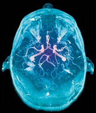
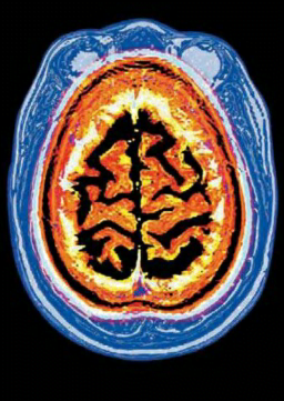
Neurologist Richard Restak likes to deliberately cloud the issue during his lectures by showing students images of two PET scans. Each reveals the level of brain activity of a student doing a problem in a Raven’s Colored Progressive Matrices test, which aims to measure “fluid intelligence,” or the ability to solve an unfamiliar kind of problem. In one scan, the image is illuminated in red , and orange, representing an increase in brain activity. In the other, the cool shades of blue and green represent a less intense level of brain function. When Restak asks the students to guess which of the two students scored higher on the Raven’s test, and thus (one assumes) possesses superior intelligence, the students invariably pick the brain lighted up like a Christmas tree. Instead, the student with the less active PET scan posted a higher Raven’s score. The explanation: The brain that finds a problem easy to solve doesn’t have to work as hard.
TYPES OF SMARTS
There are several aspects of intelligence. Most are related, but historically not all have tested what they set out to test. For example, some early IQ tests measured knowledge of facts, which actually is a function of education and memory rather than the ability to reason. In general, however, a person’s performance on a test of fluid intelligence is a good predictor of performance on a wide range of mental exercises. For example, increased fluid intelligence correlates to a high level of “working memory”-one’s ability to remember information temporarily which can range from remembering where you parked your car to which words or number combinations you tried and rejected in doing a crossword puzzle or Sudoku. People with powerful working memories are more focused in solving problems.
Scientists use the term “g-factor” when discussing the general measure of mental ability, found in vocabulary size, mechanical reasoning, and arithmetical computations. They relate it to the properties of efficient neural functioning, rather than the value of knowledge in its own right. The prefrontal cortex, right behind the forehead, is the most likely home for much of the neural processes associated with one’s g-factor abilities. When it’s damaged, a person suffers a variety of impairments to abstract reasoning, and it lights up during brain scans taken during a variety of intelligence tests.
“You have less frontal development than I should have expected,” says the evil Professor James Moriarty when he first lays eyes on Sherlock Holmes in a story by Arthur Conan Doyle. As scientists have discovered, the size of the prefrontal cortex in healthy brains generally correlates to fluid intelligence. (Perhaps Moriarty subscribed to the theory of phrenology and believed cortex size correlated to the bulging of a forehead. It’s not so.)

But the size of a cortex doesn’t mean, QED, that biology causes intelligence the same way gravity causes an apple to fall. Identical twins vary in their performance on IQ tests. In some cases, one twin develops schizophrenia or some other disorder, and the other does not. Furthermore, when identical twins are separated at birth and raised separately in similar environments, they show only a 72 percent correlation in intelligence.
FAMILY INFLUENCE
At best, genetics accounts for only a substantial fraction of intelligence. Perhaps heredity sets an upper limit for intelligence (through the potential ability to make neuronal connections), which then becomes subject to other forces. An environment with plenty of books and challenging toys plays a key role in increasing aspects of a child’s intelligence but so does willingness to exercise the brain. Political scientist James R. Flynn noted that IQ scores have dramatically increased over the past several decades in many countries. He attributes the so-called Flynn effect to increases in modern humans’ greater ability to solve abstract problems, possibly from living in a more intellectually stimulating world.
The brain’s ability to rewire neuronal networks no matter how old the nerve cells provides the means to improve mental function. Instead of looking at family or ancestral heritage and deciding it determines mental performance, humans can set about learning new skills and tasks. Challenging the brain may not raise the score on a particular IQ test, but it will help the brain to perform better.

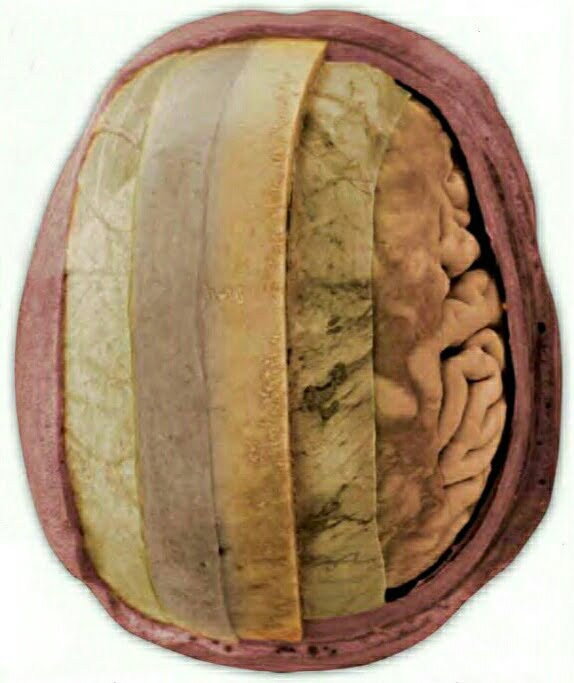
![ANATOMY [ DIFFERENT PARTS DIFFERENT RESPONSIBILITIES ( THE AMAZING BRAIN ) ]](https://humanityuapd.com/wp-content/uploads/2022/10/human-brain-vector-concept-vector-id10569719223.jpg)
![ANATOMY [ DIFFERENT PARTS & DIFFERENT RESPONSIBILITIES ( THE AMAZING BRAIN ) ]](https://humanityuapd.com/wp-content/uploads/2022/10/Screenshot_2022-10-07-12-59-20-065_com.google.android.apps_.docs_.jpg)
![NERVE CELLS - LIFE SPAN [ THE AMAZING BRAIN ]](https://humanityuapd.com/wp-content/uploads/2022/10/Screenshot_2022-10-04-13-05-57-139_com.google.android.apps_.docs4_.png)
![NEURONS AT WORK [ CONNECTIONS / ACTION / GROWTH & SUPPORT / PLASTICITY ]](https://humanityuapd.com/wp-content/uploads/2022/10/Screenshot_2022-10-02-18-44-19-068_com.google.android.apps_.docs2_-1-261x1024.png)

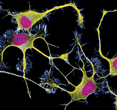
![TYPES OF GLIAL CELLS [ THE AMAZING BRAIN ]](https://humanityuapd.com/wp-content/uploads/2022/09/IMG_20220930_184056.jpg)

You must be logged in to post a comment.