Category Archives: THE NERVOUS SYSTEM [ IN HARMONY ( MANY PARTS/HEAD & BODY ) ]

GOOD FEELINGS / PLEASURE CENTERS [ THE NERVOUS SYSTEM ]

GOOD FEELINGS / PLEASURE CENTERS [ NERVOUS SYSTEM ]

GOOD FEELINGS

Pleasure also has its centers In the brain. A Tulane University neurologist stumbled across one such center in the 1950s when he tried to electrically stimulate the brains of schizophrenics to break them out of their passivity. His patients told him their implanted electrodes created pleasant sensations. The neurologist, Robert G. Heath, seized upon the results, focused his attention on the brain’s pleasure centers, and published the 1964 book The Role of Pleasure in Behavior.

Together with the discovery of pain centers in the brain, research on the physical causes of the sense of pleasure seemed to prove the ancient wisdom that humans seek to act in ways that bring them pleasure and reduce or avoid pain. New paths of investigation have led to innovative treatments for addiction, which is a form of behavior based on compulsive forms of pleasure seeking. PET scans reveal how drugs such as cocaine and heroin activate the brain’s pleasure centers. Cocaine, for example, blocks a neuron’s reuptake mechanism, which causes dopamine to linger in the synaptic cleft.

PLEASURE CENTERS

Joy, happiness, pleasure-what-ever you want to call the positive feelings that bring rewarding sensations and make life worth living-arise from the sensations of security, warmth, and social well-being combined with an awareness of the rightness of such feelings. A healthy brain recognizes the conditions that give rise to pleasure and responds to them appropriately. An unhealthy brain, or one that has learned negative behaviors such as addiction, can miss out on experiencing life’s joys. Both are primarily a matter of chemistry.

GOOD FEELINGS / PLEASURE CENTERS [ THE NERVOUS SYSTEM ]

The sensation of pleasure registers in several brain regions, including significant centers in the hypothalamus and nucleus accumbens , which lies below a portion of the basal ganglia linked to movement. All such pleasure centers rely on the chemical work performed by endorphins and neurotransmitters, particularly dopamine, to create and sustain a happy mood. Experiments with rats have demonstrated the key role of dopamine. In the 1950s, scientists wired rats’ brains so that when they pressed a bar, they received a mild electric shock to the hypothalamus. This stimulation registered as pleasure; the rats would rather press the bar than eat. However, in later experiments, rats wired for self-stimulation first received injections of drugs that block the receptors where dopamine normally binds, denying its pleasure-giving action. The rats no longer felt a pleasant reward from pressing a lever to stimulate their brain, and they stopped doing so. When humans take a similar dopamine-lowering medication, often in order to ward off hallucinations and other psychotic behavior, the drug’s success comes at a price. Delusions may leave, but so do joy and motivation. Conversely, drugs like amphetamines that increase the activity of dopamine in the brain lower the threshold for the perception of pleasure. Too much of a drug-induced pleasant sensation, however, can lead to addiction and manic moods.

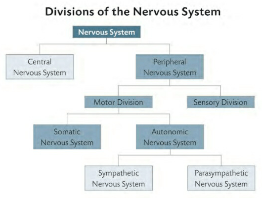
When the skin warms, the sympathetic division of the autonomic nervous system dilates blood vessels near the surface and activates the sweat glands. When body temperature cools, the autonomic nervous system narrows surface vessels to send blood to deeper, more vital organs.

“The greatest pleasure of life is love,” said the Greek playwright Euripides nearly 2,500 years ago. Like other forms of pleasure, love is processed by brain chemistry, specifically by heightened levels of neurotransmitters in the pleasure centers. MRI scans of the brain relate the feeling of lust to estrogen and androgens; attraction-more emotional than physical-appears to be associated with serotonin and dopamine. The brain chemistry that supports long-term relationships such as lifelong commitment has been harder to pin down.

Playing key roles in the sensation of pleasure are oxytocin, endorphins, and phenylethylamine , or PEA, sometimes called the love drug. These chemicals help foster the “high” felt in the first stages of love, as well as the euphoria some-times reported by long-distance runners. Even a small pleasure, such as finding your lost car keys, begins with a tiny rise of these and similar neurotransmitters in the brain’s pleasure centers.

BIG PAIN [ MESSENGERS ( THE NERVOUS SYSTEM ) ]

BIG PAIN [ MESSENGERS ( THE NERVOUS SYSTEM ) ]

It turns out, the brain has automatic defenses cued up for a quick response to more serious pain. The perception of pain warns the brain of actual or potential tissue damage. The brain’s recognition of pain sets in motion actions to reduce or remove it, and thus the threat.

Most pain receptors consist of the bare ends of sensory nerves embedded throughout all body tissues, except the brain, whose cells cannot experience sensation. These noclceptors react to any ”noxious” stimulation, anything that damages the body’s cells.

Damage makes the cells release chemicals that activate neurotransmitter receptors (substance P is the transmitter for pain) and send pain signals via the peripheral nervous system to the central nervous system, where it may take a while to be felt. Pain doesn’t reach the brain instantly because of the distance the signal must travel; in a tall man, injury to the toe may take rwo seconds to register in the brain.

In the skin, muscles, and joints, cell damage is likely to cause relatively brief and sharp pains. That’s because nerve cells in the spinal cord release natural pain suppressants known as enkephalins, which inhibit the discharge of more pain-exciting neurotransmitters and keep the sensation short. As a result, sharp pains usually fade into dull aches.

Deeper cell damage is more likely to create burns and aches that last longer. The difference lies in the kinds of nerve fibers that transmit the pain signals, and how quickly that information travels.

ASPIRIN
ASPIRIN

HIPPOCRATES, the founder of modern medicine, knew that chewing willow bark alleviated pain. Thousands of years later, scientists discovered why: The bark contains salicylic acid. When cells are damaged, they release an enzyme called cyclooxygenase-2. That chemical in turn produces prostaglan-din, which signals to the brain that part of the body is in pain. Prostaglandin also causes the injured flesh to swell and become inflamed. Salicylic acid binds to cyclooxygenase-2, blocking the creation of prostaglandin. Less prostaglandin means fewer pain signals reaching the brain, and less inflammation of the cells around the injury.


Damage to the internal organs, or viscera, usually results in dull aches, burning sensations, and gnawing pain. As the pathways for the visceral and somatic nerves of organs and body converge in the spinal cord, the brain sometimes gets confused and assigns visceral pains to other parts of the body that are not actually injured. A heart attack, for example, may seem to cause shooting arm pams.

THE AUTONOMIC NERVOUS SYSTEM / TWO BRANCHES

THE AUTONOMIC NERVOUS SYSTEM / TWO BRANCHES

THE AUTONOMIC NERVOUS SYSTEM

Much of what the brain does takes place beyond our ability to sense it-or appreciate it. In the midbrain’s pons and medulla lie the centers that regulate the vital, everyday functions of life. Think about it: How fortunate you are that you don’t have to concentrate in order to breathe, or make your heart pump blood.

The first rule of the living brain is to go on living. Thus, these crucial areas of the midbrain, called the autonomiC (“involuntary”) nervous system, are not easily overruled by the higher functions of the cortex. While it’s possible to hold your breath while underwater or throwing a tantrum, the midbrain will eventually overrule the efforts of the cortex and force the lungs to inhale. However, some drugs, such as tranquilizers and stimulants, can affect the autonomic nervous system, altering things like the heart rate and blood pressure for good or ill.

THE AUTONOMIC NERVOUS SYSTEM / TWO BRANCHES [ HARMONY ]

TWO BRANCHES

Like day and night, the autonomic nervous system has two equally important halves. They are reciprocal and complementary. The day- light side of wakefulness and work is called the sympathetic branch. It works when the body’s sense of self-preservation, developed over eons of evolution, calls for energy. In extreme cases, the sympathetic branch triggers the so-called fight or flight response. When a threat looms, the body prepares to meet it or quickly escape from it. Blood pressure and heartbeat skyrocket, breathing speeds up, and in a multitude of other ways the midbrain signals to the body to prepare itself for action.

The parasympathetic branch is the calmer, quieter side of the nervous system. It’s responsible for the so-called relaxation response. The midbrain signals to the body to lower breathing rate, heartbeat, and blood pressure. As a result, the brain promotes and recognizes a feeling of well-being.

Modern pharmacology can bring about a similar result, but much of the self-help books of the past few decades have focused on meditation and other forms of stress management to stimulate the parasympathetic branch while soothing the sympathetic.

THE CEREBRAL CORTEX [ HARMONY ( THE NERVOUS SYSTEM ) ]

THE CEREBRAL CORTEX [ HARMONY ( THE NERVOUS SYSTEM ) ]

THE CEREBRAL CORTEX

Seven-tenths of the volume of the human nervous system lies in the cerebral cortex. Given that the human cortex is many times larger than that of any other creature, scientists are convinced its huge size is the main source of what sets humans apart from the animals. Creativity, emotion, perception, language, imagination-all have strong connections to the workings of the cortex.

Beginning in the late 19th century, researchers began cataloging variations in the thickness and structure of the cerebral cortex. Korbinian Brodmann, a German neuroscientist, created a numbered map of the cortex in 1906, based on the organizational architecture of the cells that he observed after staining them. He numbered 52 sites in the brain, now called Brodmann areas. While the significance of these areas has been widely debated, further investigation has linked some of the sites to particular functions of the brain. PET scans and functional MRI scans have linked specific motor and sensory functions to specific cortical areas called domains. Brodmann areas 1, 2, and 3, for example, reside right behind the central sulcus and are closely linked to the primary somatosensory cortex, while Brodmann areas 41, 42, and 43 are associated with hearing.

The map is not a precise atlas with domains neatly separated by boundary lines, the way countries are separated by political divisions inked on paper. Many functions such as language and memory overlap domains and may in fact be scattered throughout much of the brain.

IS IT POSSIBLE to have handwriting like a serial killer’s? Does a physician’s scrawl indicate a love for humanity? Much like the phrenologists who thought a bumpy skull could reveal insights into the human psyche, so do today’s graphologists, or handwriting experts, believe that penmanship can tell us a great deal about who we are. Handwriting analysts have succeeded more than phrenologists in selling their pseudoscience. Witness the TV ads in 2008 that analyzed car buyers’ signatures. Proponents claim that because the brain controls psychological traits and muscles that produce handwriting, they must be linked. No causal link has been found. Graphologists lack scientific rigor, often analyzing the writing of people with known traits-kind of like shooting an arrow at a barn, then drawing a bull’s-eye around it.

Nor is the map an indicator of destiny, as other scientists would find. In the early 19th century, Franz Joseph Gall made his own maps of the brain and skull, but they proved faulty. He examined the bumps on the head and drew erroneous conclusions about the functions of the underlying portions of the brain. Physical variations in the size and shape of the head have nothing to do with the workings of the brain power beneath. Damage to a particular Brodmann area, however, may manifest itself in predictable ways, such as language deficiencies resulting from lesions in areas 44 and 45.

DIVISIONS [ HARMONY ( THE NERVOUS SYSTEM ) ]

DIVISIONS [ HARMONY ( THE NERVOUS SYSTEM ) ]

The peripheral nervous system has two key parts. The sensory division is sometimes called afferent, for the Latin for “carrying toward.” It sends signals from sensory receptors all over the body toward the central nervous system. Sensors in the skin, muscles, and joints are called somatic (“body”) afferent fibers, while those from the internal organs are called visceral afferent fibers.

The other part, the motor or efferent division, sends signals from the central nervous system to the muscles and glands. As these signals cause, or “effect,” changes, they create the motor responses that make the body move. Most nerve cells act as two-way streets, sending signals back and forth between the brain and extremities. Purely afferent or efferent cells are rare.

DIVISIONS [ HARMONY ( THE NERVOUS SYSTEM ) ]

The motor division also is divided into parts. The somatic nervous system sends signals from the central nervous system to the skeletal muscles. As it is usually under conscious control, this is sometimes called the voluntary nervous system. The other part is the autonomic nervous system, which comprises visceral motor fibers that automatically activate the heart, digestive tract, and other body functions.

PHRENOLOGY [ THE NERVOUS SYSTEM ( HARMONY ) ]

PHRENOLOGY [ THE NERVOUS SYSTEM ( HARMONY ) ]

PHRENOLOGY

As A SCHOOLBOY of nine, Franz Joseph Gall (1758-1828) was intrigued by a classmate with large, protruding eyes and a knack for rote memorization. The student’s appearance and skills made a lasting impression, one that years later Gall would trace to his theory of cortical localization. All the best memorizers, the German anatom ist recalled, seemed to share these bulging, “ox-like” eyes. So it followed, Gall concluded, that the function of verbal memory is governed by the frontal lobe of the cerebral cortex. The better the memory, the larger the lobe, and hence the jutting eyes.

Though he did not coin the term- and shuddered at its Usage Gall would become a leading exponent of phrenology, the pseudoscience of interpreting personal characteristics and mental abilities from cranial knobs and knots.

In interviewing hundreds of personalities across the continent and amassing a collection of some 600 skulls-not the interviewees’, fortunately-he determined the human brain to house 27 faculties. Each, he said, is controlled by different areas of the brain.

An ivory phrenological head maps skull lumps for pseudo-scientific analysis

Among those faculties we share with animals, Gall included “reproductive instinct” , “pride” , and” destructiveness, carnivorous instinct, or tendency to murder.” Unique to humans were “poetic talent,” “religious sentiment,” and “wisdom.”

Determining each faculty’s cortical coordinates was simple enough. A large percentage of pickpockets, for example, had a sizable bulge on the side of the head. This area, Gall assumed, was then location of a faculty he called “desire to possess things.” The logic of Gall’s classification system had made it widely appealing by the 1830s.

Phrenology has since been lumped with the likes of astrology, palm reading, and graphology (handwriting analysis). Yet Gall unwittingly contributed to true science. His theory of cortical localization would prompt future neuroscientists to rethink their concept of the brain, paving the way for ground- breaking discoveries at the turn of the century.

THE NERVOUS SYSTEM [ IN HARMONY ( MANY PARTS/HEAD & BODY ) ]

THE NERVOUS SYSTEM [ IN HARMONY ( MANY PARTS/HEAD & BODY ) ]

MANY PARTS

Much of what goes mto making music takes place without thought. Professional musicians don’t stop to ask themselves, How do I playa C major chord? Instead, their actions have become automatic. Likewise, some learned actions are so routinely processed that they pass out of the conscious thoughts of the cortex and are pushed deeper into the rote performance of the cerebellum.

The similarities continue. The noise of some instruments may be drowned out by the trumpets and drums, but those sounds are still there, just as the brain’s control of breathing and heartbeat continues regardless of whether they register on the mind. The conductor may step down from the podium and lower his arms; the brain rests and the body falls asleep. Or the pianist may have injured an arm and play badly or not at all, just as the signals to or from the brain may fail, and the body consequently suffers.

THE NERVOUS SYSTEM [ IN HARMONY ( MANY PARTS/HEAD & BODY ) ]

HEAD & BODY

The human body has been shaped through cephalization, an evolutionary force that concentrates nervous and sensory tissue at one end of the body. Animals under- going this process enjoy advantages in natural selection. When vision, hearing, smell, and other faculties work with a nearby brain, they provide a rich picture of the world. Specifically, having a head improves efficiency in locating food and avoiding predators.

Each division is responsible for the collection of and response to different stimuli.

A narrow gap between brain and sensory organs, such as eyes, creates the shortest pathways for information to move back and forth between the two. That reduces reaction time. Imagine the alternative: if you had organs of vision in your toes, it would take a moment longer for any images they register to reach a brain at the other end of your body, and another moment or two for the brain to send them feedback. That’s a long delay when the eyes detect a potential threat. There’s not typically a lot of variation from one head to another.

Each brain lies encased within a hard, bony skull, a series of 22 fused bones that protect it. Inside the skull is a series of protective membranes called meninges that cover the brain tissue and blood vessels, and a shock-absorbing liquid called cerebrospinal fluid. The average man’s brain weighs about 3.5 pounds; the average woman’s, 3.2. Taken as a pure ratio between brain size and body mass, that’s not a significant difference.

Like a captain on the bridge of a ship, the brain issues commands atop the spinal cord, which also lies within protective membranes, a column of bones called verte- brae, and cerebrospinal fluid. The brain communicates with most of the body through nerves that pass through the thumbwide bundle of the spinal cord inside the vertebrae, and branch out in 31 pairs of spinal nerves, each serving its own region. A few nerves, such as those that serve the face, connect directly to the brain.