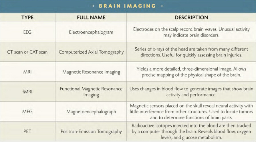
MAGNETOENCEPHALOGRAPHY ( MEG ) also relies on magnetism to examine the brain. In this case, it’s the body’s ambient magnetic fields, not those generated by an external machine, that form the basis of brain imaging. These magnetic fields are extremely weak-perhaps only a billionth of the power that causes a compass needle to point toward the north magnetic pole. Yet, when read by sensors placed on the skull, MEG scans reveal the electrical currents created by neural discharges. The resolution is as fine as a thousandth of a second and as small as a cubic centimeter. The MEG scan and EEG are the only observational techniques capable of anything approaching real-time revelations. When a patient thinks a specific thought, it shows up, in progress, on an MEG.
Mental functions also can be localized with a technique called positron-emission tomography, or PET. A radioactive isotope is injected into a patient. Because all radioactive atoms decay into stable atoms at a known rate, the decay of the isotope, which is usually paired with glucose, is recorded and turned into images with computer programs. Like MRI and CT scans, PET scans let observers localize activity inside the brain.
The array of brain-imaging techniques serves like the variety of hammers, saws, and other tools in a mechanic’s toolbox. A scientist observing the brain chooses the right tool based on what kind of information is needed. A CT or MRl scan would be the choice if a doctor suspects the growth of a tumor or physical damage to part of the cerebrum. A PET scan might be the appropriate choice for investigation of deficiencies associated with language or reason. And lack of oxygen use in stroke- damaged sections of a brain would call for a functional MRI.
A patient receives a PET scan to pinpoint regions of the brain that are most active.
True to the rational and observational methods of Descartes and Willis, science has made great strides in describing how the brain’s parts, both large and small, function. But understanding any organ that is “wider than the sky” is not as easy as toting up small pieces of information. The brain is an integrated unit, with its complexity arising from the synergy created by the simultaneous functioning of its billions of neurons and trillions of synapses in nonlinear ways. Science has learned much about movement, sensations, emotions, and the sense of self. Yet much is yet to be gleaned about the most complicated object in the universe. There will always be more to learn about the brain.
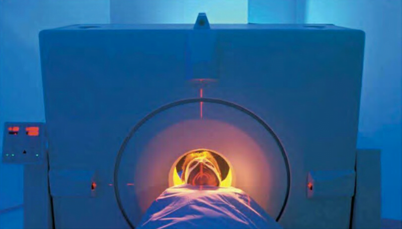
Magnetic resonance imaging (MRI) gives a more detailed 3-D picture than a CT scan. An MRI relies on an intense magnetic field generated in a cylinder that surrounds the patient. It allows precise mapping of the physical shape of the brain. Its magnetic field is so powerful that it causes some of the atoms inside the brain to jerk into alignment. Then a series of radio waves from the MRI scanner bounce off the affected atoms and push them slightly out of line. When the energy from the radio signals is turned off, the atoms move back into their magnetic alignment, emitting telltale energy patterns along the way.
Addictive drugs work by mimicking neurotransmitters or altering their work. Brain scans reveal physical changes in the synaptic activity of a drug user. The drug known as Ecstasy, for example, can permanently damage neurons that produce serotonin.
Computers read these minuscule bits of energy and assemble images of cross-sections of the brain. Slices can be placed atop each other, like the layers of a cake, to represent the entire brain in three dimensions, or they can be examined individually, providing a closer look at localized phenomena. Comparisons of MRI scans of a single brain over time can show its growth-or reveal its deterioration.
WHEN THE DENTIST asked British philosopher Bertrand Russell where he felt pain, Russell replied, with humor and honesty, “In my mind, of course.” Russell knew the brain uses the senses to collect data about the world and construct a version of “reality.” Whether that world actually exists independent of the mind makes little difference to the sufferer of a tooth- ache-the pain hurts just the same. In fact, some philosophers, such as George Berkeley (1685-1753), have questioned whether “reality” exists.
In addition to mere structure, an MRI can also capture a snapshot of thought. A variation called a functional MRI, or f-MRI, builds upon the fact that a blood cell’s magnetic properties change according to how much oxygen it contains. Receptor cells use oxygen as they take in signals from surrounding cells; burning oxygen causes cells to require more oxygen-rich blood. As blood surges toward neurons where synapses are firing with thought, emotion, or other impulses, the oxygen they carry gives off a traceable signature of radio waves. Different thoughts light up different areas of the brain in an MRI. The processes of peaking, reading, appreciating humor and music, and recognizing faces illuminate various groups of neurons. MRI techniques thus help localize areas associated with certain brain functions.
FIRST GLIMPSE / A BETTER LOOK / COMPUTERIZED VISIONS [ THE AMAGING BRAIN ]
FIRST GLIMPSE
The first technology to peer into the brain was the x-ray, invented by Wilhelm Rontgen (1845-1923) in 1895. The German scientist discovered a form of radiation that could penetrate the body; the rays were absorbed by dense bones, which then appeared as shadows on film.
When applied to the brain, simple x-rays, harnessed to make photographic images of bone, permitted doctors to make a basic examination of the structure of the head. However, x-rays give only a two-dimensional view, and show relatively little of the soft tissues of organs. As the human brain is a three-dimensional object, whatever appeared in a 2-D image usually was murky and confusing. Often, structures lying in different planes of the brain overlapped each other, making analysis difficult.
A BETTER LOOK
Scientists first peered at real-time brain functions in 1929 with the invention of the electroencephalogram, or EEG. Electrodes fitted to the scalp record electrical activity within the brain as neurons discharge. Unusual brainwave activity registered on an EEG may indicate brain disorders. This technique records electrical activity in real time.
More recently, scientists have employed a variety of tools to get a more detailed and localized look at structure and action inside the brain.
COMPUTERIZED VISIONS
Computerized axial tomograms, or CT scans, have substantially improved the ability of x-rays to probe the secrets of the brain. A patient receiving a CT scan lies inside a doughnut-shaped array of sensitive detectors while a movable x-ray emitter rotates around the brain. Computers convert the images into a three-dimensional image of the brain. Slices of the interior-the word tomos is Greek for “section”-can be teased from the data and shown on a screen to give doctors a narrow look at particular points in the brain. For example, a CT scan might reveal a tumor located deep inside the tissue of a living brain, far too deep to be visible during routine exploratory surgery.
SWEDISH SCIENTISTS in 2008 created the illusion of shaking hands with yourself. They had volunteers and a mannequin wear virtual reality goggles Images in the volunteers’ goggles came from the dummy. Most test subjects felt the weird sensation of the dummy’s point of view when shaking their own hands.
WHAT IS INTELLIGENCE [ LOOKING INSIDE ( THE AMAGING BRAIN ) ]
PERHAPS NO scientific book of the past half century stirred up as much controversy as The Bell Curve: Intelligence and Class Structure in American Life. The 1994 book, by Richard ]. Herrnstein and Charles Murray, begins simply: “That the word intelligence describes something real and that it varies from person to person IS as Universal and ancient as any understanding about the state of being human.” From there, the authors delve into definitions of intelligence and how it can serve as a good predictor for success in life.
Then they argue that different levels of intelligence lead to social outcomes, instead of the other way around a person oflow intelligence is more likely to end up a criminal or unemployed, for instance and that intelligence levels have an observable correlation to biology.
Following the track linking genetics to intelligence, the authors make claims linking racial differences to intelligence, and thus the positive and negative social outcomes that define modern life. If a group of people can’t change their biology, goes this hypothesis, they cannot change their social outcomes.
Does the brain’s biology determine intelligence, and thus lock humans in to paths toward success or failure? It’s a potent question.
DEFINING INTELLIGENCE
Part of the problem lies in the definition of intelligence. Neuroscientists don’t agree on what the word means. Nor do they agree on what intelligence tests are actually measuring. Tests don’t measure motivation, persistence, social skills, and a host of other attributes of a life that’s well lived. Some say, only half facetiously, that IQ tests measure only one’s ability to perform well on IQ tests.
Studies of identical twins have shown that certain regions of the brain are highly inheritable, affecting overall intelligence.
Neurologist Richard Restak likes to deliberately cloud the issue during his lectures by showing students images of two PET scans. Each reveals the level of brain activity of a student doing a problem in a Raven’s Colored Progressive Matrices test, which aims to measure “fluid intelligence,” or the ability to solve an unfamiliar kind of problem. In one scan, the image is illuminated in red , and orange, representing an increase in brain activity. In the other, the cool shades of blue and green represent a less intense level of brain function. When Restak asks the students to guess which of the two students scored higher on the Raven’s test, and thus (one assumes) possesses superior intelligence, the students invariably pick the brain lighted up like a Christmas tree. Instead, the student with the less active PET scan posted a higher Raven’s score. The explanation: The brain that finds a problem easy to solve doesn’t have to work as hard.
TYPES OF SMARTS
There are several aspects of intelligence. Most are related, but historically not all have tested what they set out to test. For example, some early IQ tests measured knowledge of facts, which actually is a function of education and memory rather than the ability to reason. In general, however, a person’s performance on a test of fluid intelligence is a good predictor of performance on a wide range of mental exercises. For example, increased fluid intelligence correlates to a high level of “working memory”-one’s ability to remember information temporarily which can range from remembering where you parked your car to which words or number combinations you tried and rejected in doing a crossword puzzle or Sudoku. People with powerful working memories are more focused in solving problems.
Scientists use the term “g-factor” when discussing the general measure of mental ability, found in vocabulary size, mechanical reasoning, and arithmetical computations. They relate it to the properties of efficient neural functioning, rather than the value of knowledge in its own right. The prefrontal cortex, right behind the forehead, is the most likely home for much of the neural processes associated with one’s g-factor abilities. When it’s damaged, a person suffers a variety of impairments to abstract reasoning, and it lights up during brain scans taken during a variety of intelligence tests.
“You have less frontal development than I should have expected,” says the evil Professor James Moriarty when he first lays eyes on Sherlock Holmes in a story by Arthur Conan Doyle. As scientists have discovered, the size of the prefrontal cortex in healthy brains generally correlates to fluid intelligence. (Perhaps Moriarty subscribed to the theory of phrenology and believed cortex size correlated to the bulging of a forehead. It’s not so.)
Psychologist John Raven devised the Raven’s Colored Progressive Matrices Test in 1938, a non-verbal test of intelligence in children.
But the size of a cortex doesn’t mean, QED, that biology causes intelligence the same way gravity causes an apple to fall. Identical twins vary in their performance on IQ tests. In some cases, one twin develops schizophrenia or some other disorder, and the other does not. Furthermore, when identical twins are separated at birth and raised separately in similar environments, they show only a 72 percent correlation in intelligence.
FAMILY INFLUENCE
At best, genetics accounts for only a substantial fraction of intelligence. Perhaps heredity sets an upper limit for intelligence (through the potential ability to make neuronal connections), which then becomes subject to other forces. An environment with plenty of books and challenging toys plays a key role in increasing aspects of a child’s intelligence but so does willingness to exercise the brain. Political scientist James R. Flynn noted that IQ scores have dramatically increased over the past several decades in many countries. He attributes the so-called Flynn effect to increases in modern humans’ greater ability to solve abstract problems, possibly from living in a more intellectually stimulating world.
The brain’s ability to rewire neuronal networks no matter how old the nerve cells provides the means to improve mental function. Instead of looking at family or ancestral heritage and deciding it determines mental performance, humans can set about learning new skills and tasks. Challenging the brain may not raise the score on a particular IQ test, but it will help the brain to perform better.
![BRAIN MAPPING [ LOOKING INSIDE ( THE AMAZING BRAIN ) ]](https://humanityuapd.com/wp-content/uploads/2022/10/IMG_20221014_204708.jpg)