Tag Archives: ANATOMY DIFFERENT PARTS DIFFERENT RESPONSIBILITIES

SEEING THOUGHTS [ LOOKING INSIDE ( THE AMAZING BRAIN ) ]

SEEING THOUGHTS [ LOOKING INSIDE ( THE AMAZING BRAIN ) ]

SEEING THOUGHTS

MAGNETOENCEPHALOGRAPHY ( MEG ) also relies on magnetism to examine the brain. In this case, it’s the body’s ambient magnetic fields, not those generated by an external machine, that form the basis of brain imaging. These magnetic fields are extremely weak-perhaps only a billionth of the power that causes a compass needle to point toward the north magnetic pole. Yet, when read by sensors placed on the skull, MEG scans reveal the electrical currents created by neural discharges. The resolution is as fine as a thousandth of a second and as small as a cubic centimeter. The MEG scan and EEG are the only observational techniques capable of anything approaching real-time revelations. When a patient thinks a specific thought, it shows up, in progress, on an MEG.

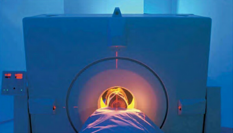
Mental functions also can be localized with a technique called positron-emission tomography, or PET. A radioactive isotope is injected into a patient. Because all radioactive atoms decay into stable atoms at a known rate, the decay of the isotope, which is usually paired with glucose, is recorded and turned into images with computer programs. Like MRI and CT scans, PET scans let observers localize activity inside the brain.

The array of brain-imaging techniques serves like the variety of hammers, saws, and other tools in a mechanic’s toolbox. A scientist observing the brain chooses the right tool based on what kind of information is needed. A CT or MRl scan would be the choice if a doctor suspects the growth of a tumor or physical damage to part of the cerebrum. A PET scan might be the appropriate choice for investigation of deficiencies associated with language or reason. And lack of oxygen use in stroke- damaged sections of a brain would call for a functional MRI.

A patient receives a PET scan to pinpoint regions of the brain that are most active.

True to the rational and observational methods of Descartes and Willis, science has made great strides in describing how the brain’s parts, both large and small, function. But understanding any organ that is “wider than the sky” is not as easy as toting up small pieces of information. The brain is an integrated unit, with its complexity arising from the synergy created by the simultaneous functioning of its billions of neurons and trillions of synapses in nonlinear ways. Science has learned much about movement, sensations, emotions, and the sense of self. Yet much is yet to be gleaned about the most complicated object in the universe. There will always be more to learn about the brain.

ANATOMY [ DIFFERENT PARTS & DIFFERENT RESPONSIBILITIES ( THE AMAZING BRAIN ) ]

ANATOMY [ DIFFERENT PARTS & DIFFERENT RESPONSIBILITIES ( THE AMAZING BRAIN ) ]

DIENCEPHALON

In the center of the brain, between the cerebrum’s two hemispheres, lies the diencephalon. It consists largely of three important structures : the Thalamus, Hypothalamus, and Epithalamus. The Thalamus acts as a relay for sensory information on its way to the cerebrum and is crucial to memory and emotions. The tiny Hypothalamus exerts control over the autonomic nervous system and performs other functions, including regulating body temperature. The Epithalamus includes the pineal gland, which drew Descartes’s attentions. Instead of housing the soul, scientists now know it helps to regulate the body’s rhythms of sleeping and wakefulness.

Elements of the diencephalon link the left and right hemispheres.
Elements of the diencephalon link THE you left and right hemispheres.

CEREBELLUM

At the back and bottom of the skull rests the cerebellum. Like the cerebrum, it too is divided into halves and deeply fissured. Its role is to coordinate movement and balance. Precise physical activities that must be practiced to be performed well-hitting a golf ball, doing gymnastics, picking a pattern of notes on the strings of a guitar-are processed in the cerebellum. The cerebellum also is known to play a role in emotion and action.

Misunderstanding of the work of neuroscientist Roger Sperry in the 1970s fed the notion that everyone is either “left brained” or “right brained.” Although each hemisphere has special functions, the two halves work closely together in a healthy mind. Humans are whole brained.

MEDULLA OBLONGATA

Where the brain meets the spinal cord is the brain stem. The spinal cord, the central route of nerve cells connecting brain and body, terminates in a 1.2 inch extension into the lower brain known as the medulla oblongata, home to motor and sensory nerves. Here is where the nerves from the body’s left and right sides cross each other on their way toward the cerebrum. Basic body functions such as heartbeat and respiration are controlled in the medulla.

Above the medulla lie the pons and midbrain. Pons means “bridge,” and that’s what it does-it acts as a bridge between the medulla and other brain regions. The midbrain links the pons to the diencephalon and controls reflexes of the ear and eye, such as the jolt the body experiences when startled.

FUELING THE BRAIN

Blood pumped from the heart pushes upward into the brain through two main sets of blood vessels, the internal carotid and vertebral arteries. Spiderwebs of smaller vessels, like distributary waterways at a river’s mouth, send blood into every region of the brain.

The brain uses oxygen III a hurry. While the brain weighs only about three pounds, a mere fraction of body weight, it burns 20 percent of the body’s oxygen and glucose. Most of that energy is mere upkeep, keeping the brain on the razor-sharp edge of action by maintaining the electric fields of the membranes surrounding the synaptic clefts. Actually thinking adds very little to the demand for energy-a fact that is somewhat counterintuitive for anyone who has ever struggled with a particularly difficult math problem or foreign language translation.

To get fuel to hungry brain cells, the body relies on the constant circulation of glucose. It’s a kind of sugar that circulates via the bloodstream. Neurons can’t stock-pile glucose like coins in a bank, so they require a ready supply of this source of chemical energy. Neurons use the fuel of glucose to manufacture and transport molecules of neurotransmitters and enzymes. They also use plenty of energy- half of the brain’s total, in fact-to transmit electro-chemical signals from cell to cell. The body obtains glucose from starches and sugars in the daily diet. Good sources include grain, fruits, and vegetables. During periods of intense concentration, glucose levels decline in brain regions associated with memory and learning. Such a decline can cause a feeling of fatigue in the body and the brain.

AN OLD BRAIN can be an amazingly healthy and creative one. Consider:

  • Ben Franklin left public service at age 82.
  • Mary Baker Eddy founded The Christian Science Monitor at age 86.
  • Robert Frost published his last collection of poems at age 88.
  • George Bernard Shaw was still writing plays at age 94.
  • Grandma Moses received a painting commission at age 99.

ANATOMY [ DIFFERENT PARTS DIFFERENT RESPONSIBILITIES ( THE AMAZING BRAIN ) ]

ANATOMY [ DIFFERENT PARTS DIFFERENT RESPONSIBILITIES ( THE AMAZING BRAIN ) ]

THE FRONTAL LOBE

A portion of the frontal lobe of each hemisphere called the precentral gyrus controls the body’s movements. Oddly, each hemisphere moves the opposite side of the body, as if the brain’s wiring some-how became crossed. Hence, the movements of the right hand and right foot, as well as the rightward gaze of both eyes, are governed by the left side of the brain. This phenomenon has been observed for centuries. Hippocrates noted that a sword injury to one side of the head impaired movement on the body’s opposite side. And while observing combat wounds during the Prusso-Danish War of 1864, German doctor Gustav Theodor Fritsch noted that if he touched the cerebral cortex as he dressed a head wound, the patient twitched on the opposite side of his body. If one hemisphere’s precentral gyrus is destroyed-during a stroke, for instance-paralysis will result in half the body.

ANATOMY [ DIFFERENT PARTS DIFFERENT RESPONSIBILITIES ( THE AMAZING BRAIN ) ]

In front of the precentral gyrus lie the premotor cortex and the prefrontal fibers. The former organizes the body’s complex physical movements, whereas the latter inhibit actions. Inhibition is useful in a variety of social settings, such as preventing shouting in a quiet movie theater.

THE BRAIN NEEDS regular exercise if its neurons area to remain sharp. Repetition of newly learned tasks helps make those new connections stronger. Without stimulation, dendrites recede and the brain settles into simpler patterns of operation. Neurologist Robert Friedland has shown that posing new challenges to the brain can help in the defense against Alzheimer’s disease.

Perhaps not surprisingly, “Use it or lose it” appears TO be true not on Iy of mental exercise but also of physical stimulation of the brain. The brain is like other organs and works better when the body is healthy. Exercising the body regularly appears to help ward off Alzheimer’s disease, as do reducing body weight, lowering blood pressure, and eating a more healthful diet. General exercise that builds up cardiovascular endurance improves blood flow to the brain. A healthy heart usually is linked to a healthy brain, especially in the brain’s “executive function, ” which is crucial to a slew of mental tasks.

A combination of physical exercise and mental gymnastics protects the brain against deterioration with age. To spur on the brain to make new neuronal connections and protect the ones it has, there are a number of activities to try, such as:
~ Learning a new language .
~ Listening to classical music.
~ Solving mental puzzles and games, like crossword puzzles and Sudoku .
~ Eating a healthful diet.
~ Walking, jogging, or cycling regularly to promote cardiovascular health .
~ Maintaining a healthy weight.

PARIETAL LOBE AND TEMPORAL LOBE

In the parietal lobe lies the somatosensory cortex, which takes in stimulations of touch and other sensations. While lower parts of the brain register pain and pressure, the sensory cortex helps localize such feelings. Damage to the sensory cortex may result in confusion about which part of the body may be registering pain.

The temporal lobe is home to the functions of hearing and appreciation of music and to some aspects of memory. Self-experience also resides in this lobe. Electrical stimulation of the temporal lobe may dredge up intense feelings from the memory-the experience of reliving the past, known as deja vu-or do just the opposite, causing familiar people and objects to become unrecognizable.

At its base, the temporal lobe connects with the limbic system, a series of brain structures also known as the animal brain. This system allows humans to experience intense emotions such as anger and fear as well as react to these feelings.

OCCIPITAL LOBE

Behind the temporal lobe, near the rear of the head, lies the brain’s visual center in the occipital lobe. Far from the eyeballs, which takes in visual information, this portion of the cerebral cortex processes electrical impulses that begin with light waves striking the retina. Wounds to the back of the head injuring the visual cortex can sometImes cause blindness.

ANATOMY [ DIFFERENT PARTS & DIFFERENT RESPONSIBILITIES ( THE AMAZING BRAIN ) ]

ANATOMY [ DIFFERENT PARTS & DIFFERENT RESPONSIBILITIES ( THE AMAZING BRAIN ) ]

FOUR DIVISIONS

Moving inward, we come to the organ itself. The brain may appear to be a Ulllform mass of folded, pink tissue. But a closer look reveals different lobes, regions, structures, and parts that all help regulate body functions, interpret information from the body, and react to stimuli. The brain has four main parts: the cerebrum, diencephalon, cerebellum, and brain stem.

  • SHAKESPEARE WEIGHS IN on the human brain in his plays:
  • “Tell me where is fancy bred, Or in the heart, or in t he head?”-The Me rchant of Venice
  • “The brain may devise laws for the blood, but a hot temper leaps o’er a cold decree.” – The Merchant of Venice
  • “Her beauty and her brain go not together. ” – Cymbeline
  • “He has not so much brain as ear-wax.” – Trai/us and Cressida
ANATOMY [ DIFFERENT PARTS & DIFFERENT RESPONSIBILITIES ( THE AMAZING BRAIN ) ]

CEREBRUM

This largest, topmost layer of the brain is the cerebrum. It’s what most people visualize when they use their brains to picture their brains. The external layer is called the cerebral cortex. Its outer por- tion is gray from the presence of billions of nerve cell bodies, while the inner portion is white from the tangle ofaxons coated in their myelin sheaths.

In 1999, scientists discovered that Albert Einstein’s inferior parietal lobe, associated with mathematical and spatial reasoning, was 15 percent wider than that of an average brain.

In the cerebral cortex lies the core of information processing that separates humans from other animals, including reason, language, and creative thought. Homo sapiens has more of its brain in the cerebral cortex-approximately 76 percent-than any other animal. (Chimpanzees rank second at 72 percent, while dolphins have only 60 percent.)

FISSURES AND HEMISPHERES

The cerebrum is divided into parts by deep fissures. The largest of the brain’s fissures is immediately evident to the naked eye. Down the center of the cerebrum, separating it into left and right hemispheres, is the longitudinal fissure. The left and right halves of the cerebrum appear to be nearly mirror images of each other.

While they look alike, the two halves perform and control very different functions. The left hemisphere long has been considered the dominant half because of its role in processing language, but the right hemisphere is gaining new attention for its role in emotions and spatial cognition, as well as the integrative function that helps bring bits of information together to create a rich image of the world.

Connecting the two hemispheres are bands of nerve fibers that allow information to be passed back and forth between the two halves of the brain. The largest bundle, containing about 200 million nerve fibers, is the corpus callosum.

Two divides known as the Sylvi an fissure and central sulcus lie on the outside edges of the hemispheres. Their locations serve as boundaries on a map, dividing the hemispheres further into four lobes. The frontal lobe lies forward of the central fissure. Between the Sylvian and central fissures are two lobes that merge together, the parietal followed by the occipital. Behind the Sylvian fissure is the temporal lobe.