The enhancement and pruning of neural networks occurs most apparently as the baby begins to develop language. Spoken languages can sound very different from each other. In all, human languages produce about 200 different spoken sounds, called phonemes. Spoken English contains just over one-sixth of those possible sounds.
A Japanese-language keyboard suggests some of the potential complexity of learning language.
Brain scans of newborns reveal that in the first few months of life, their brain recognizes the subtle differences in phonemes other than those spoken at home. Japanese infants easily recognize the difference between the sounds made by the letters R and L. However, as the Japanese language has no sound like the letter L, adults raised speaking Japanese lose their ability to distinguish it from the letter R. Similarly, English speakers learning Spanish as adults struggle to separate the subtle sounds of the letters Band P in spoken Spanish.
But babies are able to tell such differences. That’s why it’s far easier to learn a variety of languages as a child. However, as infant brains focus on processing the auditory signals of their native languages, starting at about age 11 months they lose their ability to differentiate some nonnative phonemes. Children and adults who learn new languages after having undergone “phoneme contraction” speak with an accent.
By the time a baby is three or four months of age, its behavior provides clues to its having reached new milestones in brain development. At that age, individual infants differ widely in their reaction to events and in their patterns of brain activity as measured in EEG scans.
Rs & Ls
JAPANESE WHO BEGIN studying the English language as adults struggle with the sound of the letters Rand L. It’s not the tongue that’s to blame-it’s the brain. Newborns can distinguish all phonemes, or language sounds. Between six months and one year of age, however, children lose the ability to process previously unheard language sounds. Their loss is called phoneme contraction. Since the Japanese language slurs Rand L phonemes, adults who are exposed to the separate sounds in English for the first time cannot hear, or articulate, the difference. It’s the same for English speakers learning Japanese. They can learn the words, but it’s too late for the neuronal circuits to get the sounds exactly right.
A pattern of responses known as behavioral inhibition, which includes shyness and fear when exposed to new people and experiences, occurs in one in five healthy four month olds. Their brains show higher levels of electrical activity in the right frontal lobes. Likewise, older babies who cry upon being separated from their mother have more activity in the prefrontal cortex of their right hemisphere than do children who remain calm when mom disappears from sight.
ALBERT & THE RAT
IN A 1913 manifesto, John B. Watson introduced the term behaviorism, which, he wrote, eliminated the “dividing line between man and brute” in asserting that emotions are determined not by DNA but by external stimuli. Watson built on Ivan Pavlov’s foundation of conditioned stimulus response. Foreshadowing the 1932 publication of Aldous Huxley’s novel Brave New World, Watson theorized that “man and brute” alike can be made to order. He guaranteed, for instance, to rear any of 12 random infants to take on the occupation of his choosing. Yet Watson is remembered most, perhaps, for instilling in an infant boyan irrational fear of all things white and furry.
An 11-month-old called Little Albert plays his part in a famous behaviorist experiment.
In 1919, Watson began to work with 11-month-old Little Albert, conditioning him to fear a white rat. To begin with, Albert liked his pet, trying to touch and even hold it. Watson believed this reflected a curiosity innate in all children. Later, a new stimulus was introduced: When Albert reached for the rat, Watson banged a metal bar with a carpenter’s hammer. Albert fell face-forward on the mattress, whimpering. The rat was shown repeatedly, with gong and without, until Little Albert’s congenital fear of loud noises was transferred to the rat. This phobia, Watson later learned, applied also to white rabbits, dogs, a fur coat, and even a Santa Claus mask. Presumably, Watson wrote, Albert could eventually become unconditioned, but the boy was adopted before further experiments could be performed.
Some scientists argue that as the brain incorporates new experiences and makes new connections among neurons, it expresses a form of evolution through the competition of its various neural networks. Nobel Prize-winning neuroscientist Gerald Edelman suggests that the brain’s many networks vie against each other in “neural Darwinism.”
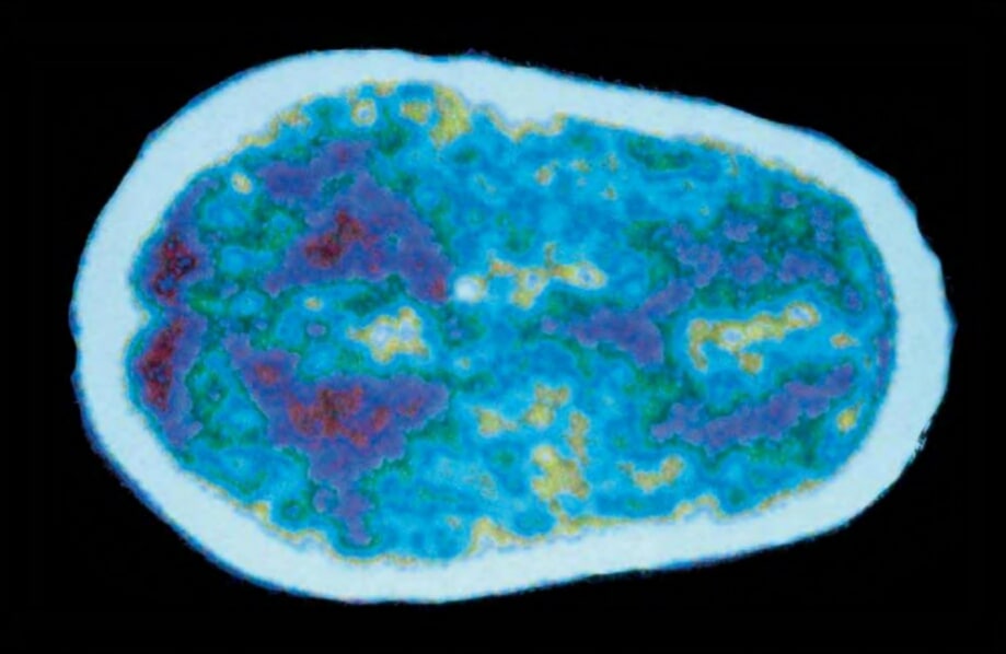
A newborn’s brain (seen above in an MRI) is ready to begin making, remaking, and pruning neural connections by the million.
While genes determine how the brain begins to grow in an embryo, the brain’s extreme complexity and plasticity make it nearly impossible to predict how it will develop in response to a particular stimulus. The complexity of the brain makes it like the weather. Short-term weather forecasts are possible with some degree of confidence, but long-range forecasts become more and more difficult because of the interaction of so many variables. The so-called butterfly effect, which was discovered during computer generated weather simulations in the 1960s, posits that under the right conditions, the flapping of a butterfly’s wings in China can be magnified until it causes a tornado in Texas. As expressed in the brain, a small change in biochemistry under sensitive conditions may have a tremendous impact on the brain’s future development.
PREMATURE births pose special challenges to the brain. The child emerges from the womb before its neural networks have been established and have gone through initial stages of pruning. Much of the brain development must occur in the buzzing confusion of the world rather than a calm womb, which psychologist Sigmund Freud called the baby’s stimulus barrier. Development of the preemie’s brain occurs without the nutrients and protection of the uterine environment. In addition to difficulties involving regulation of body temperature, digestion of food, and weakened breathing, many preemies suffer brainhemorrhage. Babies who survive amid the chaos of lightsand sounds in a hospital nursery may have their brain overstimulated and may develop problems such as attention disorders and learning disabilities later in life.
Brigham and Women’s Hospital in Boston has attempted to re-create the conditions of the womb in its neonatal intensive care unit. A preemie’s brain reacts with extreme sensitivity to light and loud noises, so the hospital keeps its NICU dark and quiet. Babies get plenty of skin-to-skin contact, to mimic the touch of the womb. They feed on demand. And they’re allowed some freedom of movement, as they would experience inside the womb, rather than being swaddled tightly The result: These babies leave the hospital earlier than those raised in a standard intensive care unit and have an accelerated developmental curve compared with other preemies.
Consider how neural Darwinism finds expression in the early stages of fetal brain growth. Neurons forming from stem cells move through the brain, guided by basic genetic coding. Genes determine how the neurons connect, axon to dendrite, to create the foundation and basic architecture of the brain. However, the precise chemical environment surrounding the newly formed neurons strongly influences how far they migrate and which neighboring neurons they link with. Exposure to substances in the womb, such as alcohol, can disrupt neuronal migration, but there is no guarantee that exposure will or won’t lead to fetal alcohol syndrome. The unpredictability of the complex system that is the human brain makes such precise calculations impossible.
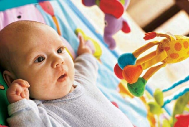
Toys and a mentally stimulating environment help a baby’s brain grow complex neural connections.
Babies don’t learn to walk until about a year after birth, but they are born with the neural program already hardwired.
As people grow older, they take in new experiences. There may be changes in climate, social networks, formal education, and career. To get on in life, people have to adapt to change. Successful adaptation is a matter of rewiring the brain by creating new neuronal connections. Links that promotesurvival and well-being grow stronger. Those that lose their usefulness grow weaker. In a process that resembles natural selection, they lose the competition to stronger neural networks, and they die.
Neural Darwinism provides a new perspective on the brain’s plasticity: As neural networks compete, those that function best get stronger. Changes in the environment encourage changes in the brain by giving new neural networks a chance to flourish. Such evolution of a single brain continues over an entire lifetime.
The most dynamic growth occurs in the cerebral cortex, the largest and outermost layer of the brain.During the first months of fetal development, when 250,000 new nerve cells are being created every minute, neurons begin to take on specialized tasks.
First, they inch their way from where they were formed by cell division to their permanent home in other regions of the brain. Most go toward the cortex, but some move into the cerebellum and other portions of the brain. This process, known as migration, is quite remarkable for the distance the neurons must travel as well as their ability to navigate surely along the tangled pathways of the developing brain. Millions of neurons migrate a distance equivalent to a person hiking from Los Angeles to Boston. Amazingly, they manage to arrive at Paul Revere’s house, the U.S.S. Constitution, or Faneuil Hall without ever consulting a map.
Once the migrating neurons reach their destination, they developed axons and dendrites to reach out and make connections with other neurons. Like roads that connect to create a grid for traffic, neurons set up systems of communication that link all parts of the brain. Some pathways receive huge amounts of sensory traffic and become the equivalent of information highways. Others turn into dead ends or decay into crumbling blacktop from lack of use.
You can’t clone a brain. And even if you could, it wouldn’t turn out like the original. Sensitivity to initial conditions in the womb coupled with differences in environment after birth would significantly alter development despite the identical genetic code.
UNDERSTANDING MIGRATION
The brain reacts with extreme sensitivity to anything that influence neuronal migration. Only a few decades ago, neuroscientists believed that each neuron had its own special, predetermined location when it set out on its trek across the brain. Now, researchers have found that neurons take on different characteristics because of their journey and their destination. To take just one example, neurons that process oral communication are not inherently preprogrammed to be speech neurons. Instead, they become speech neurons by migrating to the areas of the brain associated with language.
This discovery prompted new understanding of a wide variety of brain disorders. If something interferes with neurons migrating to their intended destinations and not overshooting or undershooting their targets the results can be powerful. Such disorders as autism, schizophrenia, dyslexia, and epilepsy have been at least partly linked with abnormalities in neuronal migration.
Fetal alcohol syndrome has also been linked to problems in migration. The brain’s hypersensitivity to toxins that impede migration underscores the warnings given to expectant mothers to avoid exposing a developing baby to alcohol, tobacco smoke, drugs, or other chemicals that may interfere with healthy brain development.
WHEN SPERM meets egg, the merger of a father’s and mother’s DNA triggers the start of a new life. Encoded in the tens of thousands of genes that make up a human being are a substantial fraction that will create the brain and central nervous system. You won’t find the child’s personality, emotions, and ideas buried in the code; they arise instead as the brain develops and interacts with its environment after birth. Nevertheless, the explosion of cell development that begins with conception is the first step toward forming the brain and all of the hopes and dreams it will one day contain.
As an embryo develops into a fetus, the brain grows and differentiates rapidly.
DIVISIONS & LAYERS
In its first phases of development, the fertilized egg, or zygote, undergoes a rapid series of divisions. One cell becomes two, two become four, four become eight, and so on until the exponential divisions Create a tiny, hollow ball of hundreds of cells nearly uniform in design.Two weeks after conception, the sphere of cells, still dividing, takes the first step in the series of physical changes to construct a differentiated body and begin the process of becoming human.
First, a dent appears in the sphere. Cells move into the indentation, which folds under the surface of the sphere. The folding creates three layers of cells: an outer layer called the ectoderm, an inner layer called the endoderm, and a middle layer called the mesoderm. In the following weeks, these three layers grow into the tissues that give rise to the body’s major systems: Endoderm becomes digestive tract; mesoderm creates muscles, skeleton, heart, and genitalia; and ectoderm forms brain, spine, nerves, and skin.
Lots of gentle handling produced increased serotonin, a neurotransmitter that dampens aggression, in baby rats. Grown into adults, the rats lived longer and handled stress better.
BUDDING BRAIN
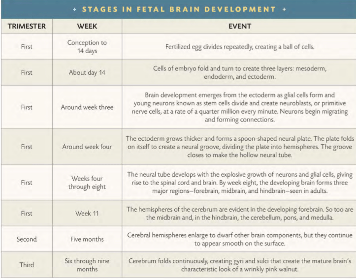
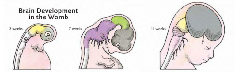
The nascent brain makes its first appearance at about four weeks after conception, when a thin, spoon-shaped layer of cells called a neural plate emerges at the head end of the embryo. Major characteristics of the future brain already are in place just one month into fetal development. Hemispheres later will develop on either side of a groove down the center of the neural plate, creating the bilateral symmetry of the human brain.
As the fetus grows, the bowl of the spoon will become the brain itself, while its handle grows into the spinal cord. And as the neural plate folds to form a tube, swellings in the original spoon shape become the forebrain, midbrain, and hind brain. As they develop, they work together to form the major sections of the brain, from the cerebrum at the top of the head to the thalamus, hypothalamus, cerebellum, and spinal cord at the back and lower end.
As modern humans evolved from their hominid ancestors, their brain development continued with increasing specialization of regions and functions. One hypothesis suggests that the differences between the left and right hemispheres of the human brain can be traced tohumans’ simian ancestors swinging through trees. Grasping one limb after another requires the arms to act independently instead of in unison. Perhaps the ancestors of humans began emphasizing the use of one arm over another, encouraging greater neuronal development in the hemisphere that controlled action on that side of the body.
One of the most pronounced differences between brain hemispheres can be observed in dissection of cadavers. The brain region mainly responsible for speech, the planum temporale, is larger in the left hemisphere of two-thirds of human brains. The left-handed nature of language is evident across time and stage of life. Full-term fetuses exhibit larger, speech-related regions in the left hemisphere than in mirror locations on the right hemisphere. The same was true of Neanderthals, according to the telltale marks on the inside of their 50,OOO-year-old skulls made by contact with their gyri and sulci.
GENDER DIFFERENCES
The two sexes also experience differences in brain function. Men are more likely to be left-handed, dyslexic, hyperactive, and autistic. Women are more likely to suffer migraines and, on average, have weaker spatial functioning. Women, though, generally outperform men in the fine motor skills of their fingers, and they learn to speak their native language earlier and foreign languages more easily than men. The bottom line, however, is that if you were to look at two brains on a laboratory table-one from a man, and the other from a woman-you probably wouldn’t be able to tell any difference.
In men, the third interstitial nucleus of the hypothalamus typically is twice as big as it is in women’s brains. The hypothalamus is crucial to sexual behavior, as well as regulation of body temperature, eating, and drinking. Furthermore, women’s and men’s brains differ in response to orgasm. PET scans show less activity in a woman’s prefrontal cortex and in a man’s amygdala during sexual climax, while both sexes experience more neuronal firing in the cerebellum.
GENDERED BRAIN
THE SEXES DIFFER in cognitive ways. A big one involves spatial orientation. Men typically use mental maps, while women prefer landmarks. Men would likely give directions by saying, “Drive north 2.2 miles, turn east, and drive 1.5 miles,” whereas women would more likely say, “Drive toward the mountains until you see the barn, turn right, and go to the pond.” Small wonder that one sex may get frustrated giving directions to the other. Women take the prize for remembering objects’ locations-where are those keys?- while men win at abstract spatial reasoning, such as mentally rotating objects. As a group, men have a wider dispersal of scores on some mental tests.
PREPROGRAMMING
Much human behavior arises from culture and environment. Some, however, appears to be prewired into the brain. The capacity for language appears to be so strongly encoded that children raised without exposure to any language will make up their own.
Communication is an evolutionary favored social activity that helps humans compete with other animals for resources necessary for life. Similarly, the brain’s ability to process and integrate visual stimuli exists almost immediately after birth. At only a few weeks old, an infant raises its arms to protect itself from the approach of an object. Sight, texture, and size appear to be aspects of object recognition that the brain is prewired to bring together for self-defense.
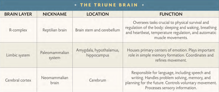
Neuroscientist Paul MacLean suggested in 1967 that the human brain functions as three separate “brains,” each of which represents a stage in evolutionary development. He referred to the three-way unity as humanity’s triune brain. Through evolution’s penchant for preserving genetic code that proves useful for survival and discarding mutations that prove useless, MacLean suggested that human brains evolved by adding to successful brain structures of earlier vertebrates. Thus, both fish and dogs have brain structures in common with people. But instead of the evolutionary structures being uniformly mixed throughout the human brain, they nest one inside another like Russian dolls. The most primitive lies deepest in the brain, under more modern layers.
Charles Darwin observed that domesticated animals have thinner cortical layers than their wild cousins in the forest. Wild animals’ exposure to a wider variety of environmental stimuli may create richer neural connections.
FIRST BRAIN
MacLean’s first “brain” is the R-complex, which takes its name from its resemblance to the simple brains of reptiles. The R-complex formed from an extension of the upper brain stem. It’s enough to keep a snake or a salamander alive as well as ensure the continuation of the species. The R-complex oversees sleeping and waking, breathing and heartbeat, temperature regulation, and automatic muscle movements. It also plays a crucial role in the processing of sensory signals from the peripheral nervous system. MacLean’s experiments with a variety of animals demonstrated that the neural connections in the R-complex provide sufficient mental firepower for hunting, mating, establishing territory, and fighting. In other words, everything necessary for finding food, competing with other animals for survival, and passing along the genes of the dominant, strongest individuals. Humans may think of themselves as being far above turtles and alligators, but their brain shares the same mechanics for regulating basic body functions. Further-more, whenever humans engage in a schoolyard scuffie or compete for the affections of another, they’re exercising the reptilian cores of their brain.
SECOND BRAIN
The second “brain” is the limbic, or paleo mammalian, system. It’s common to all mammals, including humans, but is lacking in reptiles. The limbic system coordinates and refines movement. It gives rise to emotions and simple memory, as well as the rudimentary social behaviors they make possible. When MacLean destroyed part of the limbic system in the brain of young mammals, their behavior regressed toward the reptilian. They stopped playing and exhibited weaker mother-offspring bonds. Humans who flush with anger when they get slapped across the face, or glow with happiness when kissed, are using their limbic systems. If they choose to ignore the slap or the kiss, however, they need to exercise the third and highest level of the brain.
Swinging through forest has been linked in theory to brain hemisphere specialization.
THIRD BRAIN
The third “brain” is the cerebral cortex. Many mammals possess a cortex, but it is most highly developed in humans. It adds the benefits of problem solving and both long-term and complex working memory to the lower two “brains.” The neomammalian brain, as MacLean dubbed it, gives humanity its capacity for language, culture, memory of the past, and anticipation of the future. It also makes humans the first species with empathy, the ability to see the world through the eyes of others.
“It is this new development that makes possible the insight required to plan for the needs of others as well as the self … In creating for the first time a creature with a concern for all living things, nature accomplished a 180-degree turn-about from what had previously been a reptile-eat-reptile and dog- eat-dog world,” MacLean said.
GROWING COMPLEXITY [ EVOLUTION ( BRAIN DEVELOPMENT ) ]
GROWING COMPLEXITY
If 2,000 neurons are sufficient for simple learning, imagine the explosion of complex behavior that accompanied the growth of neural complexity about 530 million years ago. Larger clumps of neurons in the diverse animal population that seemingly emerged overnight encouraged the flourishing of new animal species. The variety of new species could better react to, and survive, changes in their environments. Ocean life diversified into the ancestors of today’s worms, mollusks, and crustaceans.
The forward tip of the neural cords in the first proto-vertebrates began swelling and folding to create primitive brains. Neural networks in those early brains began to diversifY. Some connections began to specialize in vision. Some took on the function of hearing. Among the sharks, neural connections specializing in smell became hypersensitive, empowering them to detect blood in concentrations as small as 1 part per 25 million of water. That allowed them to smell bloody prey a third of a mile away (and, not coincidentally, strengthened their chances for survival in the constant interspecies combat of evolution).
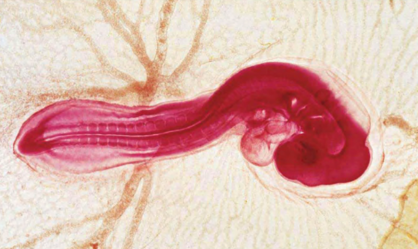
A developing spinal cord is already visible in a three-day-old chicken embryo developing inside its eggshell.
As animals began crawling out of the ocean onto the shore, around 360 million years ago, their brain didn’t begin anew. Instead, new experiences and new evolutionary developments were laid down atop their existing neural networks. Birds and reptiles added new levels of behavior, and new brain matter developed as well. Mammals put their own layers on top of their evolutionary predecessors. And finally, humans with their gigantic brain added the newest and most complex layers in the wrinkly pink walnut of the cerebral cortex.
Darwin explicitly put humans in the crosshairs of his theory with the 1871 publication of The Descent of Man. Human bodies and brains evolved and continue to do so.
The human brain differs physically from those of other mammals in its size, complexity, and dominance of its cerebral cortex. Just like speed and strength, early advantages in the brain such as analytical power (“How can I trap that animal?”) and capacity for speech (“How can I get others to help me trap that animal?”) improved the odds of early humans’ survival. Advantages spread to new generations and became common.
Networks of synapses constantly compete with each other; roughly like animal species fighting for limited food. Networks that get steady stimulation grow stronger; while others atrophy. Nobel laureate Gerald Edelman calls the process neural Darwinism.
EVOLUTION – GROWTH & ADAPTATION OF THE HUMAN BRAIN [ BRAIN DEVELOPMENT ]
FROM THE single celled product of conception, the human animal grows into a complex, uniquely cognitive being. Evolution has built upon older, more primitive animal brain forms to lead humanity to emotion and rational thought. Over eons of time, neural circuitry has developed to promote and continue to promote individual and collective survival. That’s because the human brain is “plastic,” primed from an extremely young age to learn and change.
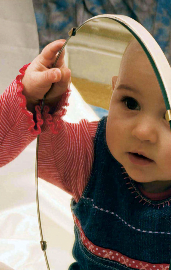
A six-month-old girl examines her reflection. From birth, humans appear to be drawn toward faces.
EVOLUTION
THE DEVELOPMENT of the human brain is written in millions of years of evolution, its story still unfolding.
Neurons began to emerge with the appearance of multicellular animals. The earliest neural connections formed primitive networks of cells in tiny life-forms swimming in primordial oceans. Today, such systems can still be found in simple life-forms such as jellyfish.
SIMPLE BRAINS
Animals with only the barest collection of neurons can function with surprising sophistication. The marine snail Aplysia has only about 2,000 neurons, yet it is capable of movement, reaction to touch, sensation, and all of the things that make a snail live like a snail. It even can learn despite lacking a true brain. Aplysia’s neurons organize themselves into clumps called ganglia at various points on its tiny body, creating a maze of connections. These neural clumps can amplifY or tamp down electrochemical signals as they pass from neuron to neuron; its neural connections can be strengthened or weakened just as in human brains. Scientists have found that when they shock Aplysia’s tail, it reacts by reflexits neural network contracts the affected flesh to pull it away from the source of the shock. However, things get interesting when the shock is preceded by a light touch against the snail’s flesh. After a few repetitions, the lowly Aplysia has enough neural complexity to connect the two sensa- tions: touch, followed by pain. In time, the light touch alone, with no electric shock afterward, is enough to make the snail recoil as if in pain.
An octopus’s brain is dime size, but it can solve simple problems such as moving barriers to get food.
CHARLES DARWIN KNEW he had opened a tinderbox when he published On the Origin of Species in 1859. He laid out a theory of evolution through natural selection: Individuals that have a biological advantage are more likely to outlive their peers and pass their edge to offspring. A gazelle that is a bit faster than another may outrun the lion and breed fast children the next day. Cuidado, Darwin wrote in his notebook, using the Spanish for “careful.” Taken toits logical conclusion, even humans fell under his theory-an idea Darwin down-played at first because he knew it would be unpopular.
The nervous system isn’t the only method by which the brain controls the body and maintains homeostasis. The direct, electrochemical means by which the nervous system collects information from stimuli and then formulates responses is augmented by the endocrine system, which works with the nervous system to regulate the body’s cells. The autonomic nervous system responds to changes in the body’s dynamic balance by releasing electrochemical impulses to the body’s endocrine organs. These include the testes and ovaries, pancreas, adrenal glands atop the kidneys, thymus and parathyroid glands, and three glands in the brain: the pineal, hypothalamus, and pituitary.
Endocrine glands respond to the nervous system’s orders by releasing hormones into the bloodstream. Hormones (from the Greek for “to excite”) bind to specific cell recep- tors and affect virtually every cell in the body. For example, instructions from the brain, given at the proper time, order the endrocrine glands to release the hormones responsible for sexual development to trigger puberty at adolescence. Other hormones maintain the body’s balance of energy, keep the blood’s supply of electrolytes in balance, and muster the immune defenses against infection. The nervous system and the endocrine system share a special relationship, as their functions can seem intricately intertwined.
FOR A HEALTHY BRAIN, good foods are a key part of optimizing your brain’s performance. Here are some foods your brain will welcome:
Fresh fruits and vegetables. These include blueberries, leafy vegetables, broccoli, and cauliflower. They contain high amounts of acetylcholine and useful vitamins. Certain vitamins, notably vitamins C and E, and beta-carotene, a precursor to vitamin A, act as antioxidants. They neutralize destructive molecules and atoms known as free radicals, which damage brain cells by stealing electrons from cellular molecules or atoms.
Unsalted nuts. Their omega-3 fats help keep the brain and nervous system healthy. Neurons require fats in their myelin sheaths to function properly.
Fish. It’s a better source of proteins than high-fat meat, and it’s another source of omega-3 fats.
Chicken without the skin and lean meats. Protein in the meat helps build tissue and supply the amino acids that form neurotransmitters .
Fruit juice. It’s a natural source of beneficial vitamins, including antioxidants. Be sure to drink plenty of water, too, to keep your brain and body hydrated .
Small amounts of alcohol, such as one glass of wine a day. This may increase blood flow to the brain and lower the risk of strokes .
Small amounts of caffeine. It activates the cerebral cortex and helps release the neurotransmitter epinephrine .
Pasta, cereal, and bread. They contain carbohydrates for energy as well as being rich in serotonin.
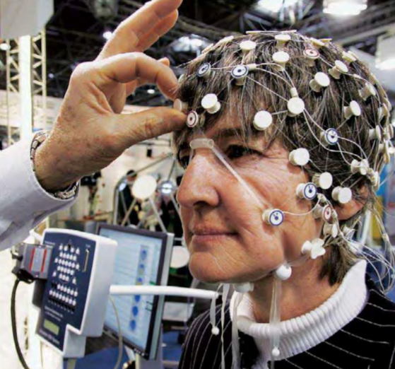
On a summer day, storm clouds can suddenly gather and transform an afternoon of sunshine into a violent monster of rain, hail, lightning bolts, and the occasional twister. Sunlight and warmth get blotted out. So it is with the nervous system. The brain’s higher functions, working in harmony with the body, promote consciousness and a sense of well-being. But because the brain functions through the medium of electrochemical reactions, the occasional storm knocks the brain out of balance.
A woman wears an EEG sensor net that aids in epilepsy analysis.
Epilepsy is a flood of electrical discharges in groups of cranial neurons. While the brain suffers through its own electrical storms, no other signals get passed through. Those who suffer an attack may fall to the ground, black out, foam at the mouth, and jerk about uncontrollably. Epileptic seizures can last from a few seconds to a few minutes, and can vary widely in their ferocity.
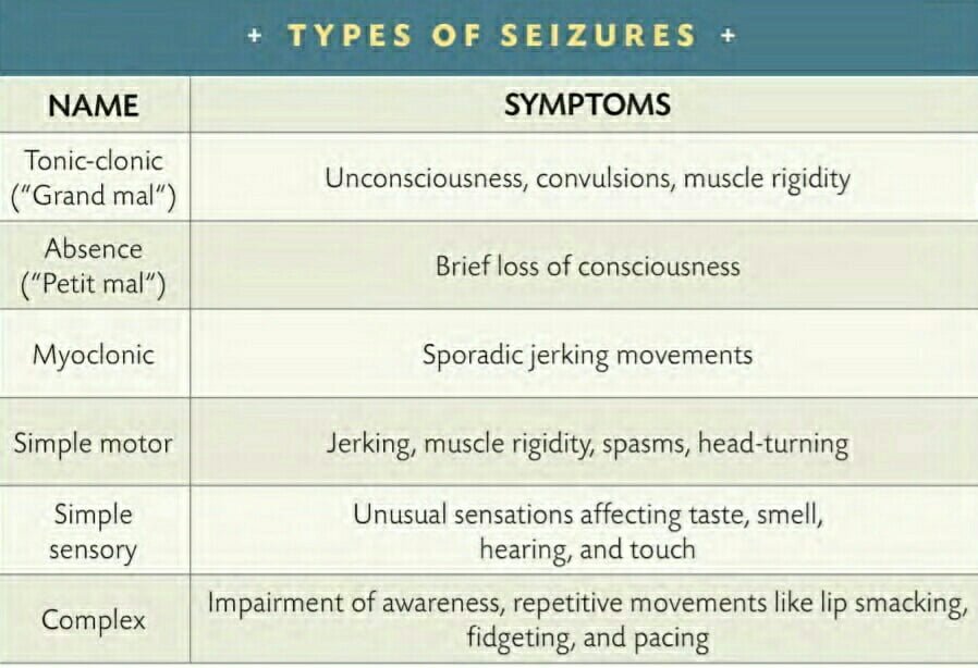
TYPES OF EPILEPTIC SEIZURE
The mildest used to be called petit mal, French for “little illness.” Now they’re referred to as absence seizures. Sufferers, usually young children, lose consciousness for a few seconds, often staring blankly into space. They typically do not know what has happened to them. Such seizures usually go away by age ten.
Stronger, convulsive seizures are called tonic-clonic, which replaces the old term, grand mal, French for “big illness.” Epileptics in the midst of a tonic-clonic seizure lose consciousness and may experience loss of bowel or bladder control, as well as muscle contractions so severe they have been known to break bones. After a few minutes, when a major seizure dissipates, thesufferer slowly regams awareness. Some tonic-clonic attacks give fair warning. Sensory hallucinations known as auras, including smells and bright lights, give the sufferer a chance to lie on the floor before the onset to avoid the potential injury of falling.
DIVINE ILLNESS
A NEUROSCIENCE JOURNAL article in 1997 listed religious figures thought to be linked with epilepsy because of recorded accounts that match its symptoms. The historical figures included:
Saint Paul, apostle and writer of much of the New Testament.
Joan of Arc, 15th-century saint and heroine of France.
Emanuel Swedenborg, 18th-century theologian.
Ann Lee, 18th-century leader of the “Shaking Quakers,” or Shakers.
Joseph Smith, 19th-century founder of the Church of Jesus Christ of Latter-day Saints, commonly called the Mormon Church.
CAUSES & TREATMENTS
Epilepsy has a variety of causes. Some are genetic in origin and caused by an inherent problem in the brain. Typically, the disease strikes far more men than it does women. Other cases have their onset after physical injuries to the brain, such as strokes, fevers, tumors, or head wounds.
About the size of an almond, the small hypothalamus plays a big role in both the nervous and endocrine systems.
Treatment options include anticonvulsive drugs and vagus nerve stimulation. In the latter, stimulators are implanted in the chest to send regular pulses of electricity through the vagus nerve to the brain. These pulses aim to keep the brain’s electrical activity from tipping from order to chaos.
New possibilities include the implantation of monitoring devices combined with electrical stimulators or drugs. The idea is to detect the subtle electrical changes that signal an oncoming epileptic seizure, then deliver a small shock or dose of medicine to ward off the attack before it strikes.
Epilepsy is an ancient disease that has fascinated and frightened scientists and laymen alike. Before we acquired a working knowledge of the central nervous system, seizures were shrouded in mystery. In antiquity, the disease was accredited to gods and demonic possession, causing those with epilepsy to be feared and isolated. Epilepsy patients continued to face discrimination through the mid-20th century. This discrimination ranged from lack of access to health insurance, jobs, marriage inequality, and even forced sterilizations. Despite the strides that have been made, there are still many misconceptions globally regarding epilepsy. While there has been substantial progress, more work needs to be done to educate people across the globe about the pathology of the disease, its causes, and mechanisms. Studies show that patients with epilepsy living in communities that understand the pathology and cause of seizures are generally more successful in social and educational environments. In this book, beyond current treatments that may include anti-epileptic drugs (also called anti-seizure medications), neurosurgery, neuro-stimulation, lifestyle modifications, and dietary changes, I ( Author ) will discuss the recent modalities of gene therapy, immunotherapy, and neutrophil therapy, and will outline more advanced research options, some of which remain to be pursued. I ( Author ) will also posit that the root cause of epilepsy is an autoimmune disease that had gone rogue, damaging the brain’s normal functions and leading to neurodegenerative diseases, including epilepsy. Under this theory, the seizures are but the symptoms of that disease. Brain function being highly non-linear, it is not too surprising that anti-seizure/anti-epileptic drugs that assume a linear brain function have been only partly successful. In all these endeavors, the well-being of the patient is foremost, and that is why I ( Author ) will also include suggestions, recommendations, and available supporting resources for patients and their caregivers, how they can live and cope with their epilepsy, and what they can do about it.
About the Author
DR. ALAIN L. FYMAT is a medical-physical scientist and an educator. He is the current President/ CEO and Institute Professor at the International Institute of Medicine & Science with a previous appointment as Executive Vice President/Chief Operating Officer and Professor at the Weil Institute of Critical Care Medicine, California, U.S.A. He was formerly Professor of Radiology, Radiological Sciences, Radiation Oncology, Critical Care Medicine, and Physics at several U.S. and European Universities. Earlier, he was Deputy Director (Western Region) of the U.S. Department of Veterans Affairs (Office of Research Oversight). At the Loma Linda Veterans Affairs Medical Center, he was Scientific Director of Radiology, Director of the Magnetic Resonance Imaging Center and, for a time, Acting Chair of Radiology. Previously, he was Director of the Division of Biomedical and Bio-behavioral Research at the University of California at Los Angeles/Drew University of Medicine and Science. He was also Scientific Advisor to the U.S. National Academy of Sciences, National Research Council, for its postdoctoral programs tenable at the California Institute of Technology and Member of the Advisory Group for Research & Development, North Atlantic Treaty Organization (NATO). He is Health Advisor to the American Heart & Stroke Association, Coachella Valley Division, California. He is a frequent Keynote Speaker and Organizing Committee member at several international scientific/medical conferences. He has lectured extensively in the U.S.A., Canada, Europe, Asia, and Africa. He has published in excess of 525 scholarly scientific publications and books. He is also Editor-in-Chief, Honorable Editor or Editor of numerous medical/scientific journals to which he regularly contributes. He is a member of the New York Academy of Sciences and the European Union Academy of Sciences, a board member of several institutions, and a reviewer for the prestigious UNESCO Newton Prize, United Kingdom National Commission for UNESCO.
MAPPING SEIZURES [ DELICATE BALANCE – THE NERVOUS SYSTEM ]
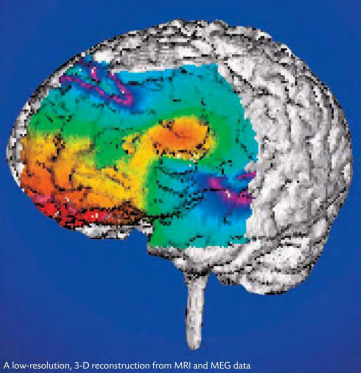
Seizures may occur in any part of the brain; their point of origin often can be mapped. Some occur as a result of lesions in specific domains. Nineteenth-century doctor John Hughlings Jackson, an aloof but meticulous researcher, posited that lesions would produce two effects. He based this belief on the idea that most of the neurotransmitters in the brain at any given moment inhibit action. A minority of neurons at anyone time release neurotransmitters that bind to receptors. Others do nothing. Thus, Jackson said lesions would produce negative reactions because of the destruction of brain tissue. However, they also would have the opposite reaction of freeing other, healthy areas of the brain, which previously had been suppressed.
The minus and plus aspects of brain damage appeared to match the observed effects of a brain tumor in a teenage girl named Bhagawhandi in the 1970s. A neuroscientist who observed the girl diagnosed a malignant brain tumor. As the tumor grew to press on her temporal lobe and her brain started to swell, she suffered a series of seizures. They grew more frequent. However, whereas her initial seizures were intense grand mal convulsions, her new manifestations, localized in the temporal lobe, were weaker. She began experiencing dreamy states in which she saw visions of her home in India. Far from being unpleasant, they made her happy-“They take me back home,” she said. She remained peaceful and lucid during her episodes. The seizures killed her in a few weeks, but doctors often noted the rapt expression on her face as she moved deeper into her visions. Only a few diseases of the central nervous system produce pleasure. Anything that pushes the brain out of homeostasis is more likely to bring pain and discomfort to the body.
A photomicrograph of L-dopa, suggestive of an abstract painting, hints at the complex world of neurochemistry.
The beauty of L-dopa lay in aseemingly simple but startling idea for treatment: If the neurons’ ability to make dopamine had dramatically decreased, why not merely supplement the supply of the drug in the brain? Not only did L-dopa help the encephalitis lethargica patients, it also became a popular treatment for a far more common disease, Parkinson’s disease, marked by muscle rigidity and loss of motor control.
Despite its ability to ease suffering, though, L-dopa is no “magiC bullet,” no magic cure. Sacks’s patients began relapsing into their former patterns of tics and frenzies. Parkinson’s sufferersalso found that over time, L-dopa lost some of its power to help them. Still, the tangible results of L-dopa treatments have encouraged neuroscientists to seek the right combination of medications to restore balance to brain chemistry for a variety of illnesses.
SEIZURES [ DELICATE BALANCE – THE BRAIN’S EQUILIBRIUM ( THE NERVOUS SYSTEM ) ]
Abnormal electrical activity in the brain produces seizures, which have a broad range of manifestations. Some are so minor that they may occur unnoticed, while others can cause violent spasms and convulsions. Victims may even lose consciousness. They can be a one time event or occur frequently.
A number of things can cause seizures: Serious conditions like strokes, brain tumors, and severe head injuries can generate them, as well as other seemingly harmless things like bright, rapidly flashing lights and low blood sugar.
There are two general types of seizures: generalized and partial. Generalized seIZures involve both sides of the brain from the beginning of an episode while partial seizures begin in specific regions of the brain and may spread to the entire brain. Generalized seizures have several subtypes, from tonicclonic seizures (formerly known as grand mal) to absence seizures (also known as petit mal).
FIRST THEY felt hyperactive and frenzied. Then their body motions became more violent, and they would twitch and convulse. Finally, they fell into a deep trance. And there they remained, these sufferers of the disease encephalitis lethargica, until neuroscientist Oliver Sacks found them in the 1960s-40 years later. As depicted in the movie Awakenings (1990), Sacks gave them L-dopa, which the brain transforms into dopamine. The dopamine levels in the postencephalitic patients had been greatly diminished by their disease. The patients woke up from their stupor, and health seemed to be restored to them.
THE DAMAGE caused by headaches is eye-popping. About 45 million Americans suffer them regularly, and about half of the sufferers find the pain severe and sometimes disabling. The result: lost time from work, play, the day to day stuff of life. Counting only the 30 million who suffer migraine headaches one of the 150 described categories of headaches American victims lose 157 million work days each year.
ALL IN YOUR HEAD?
Victims often describe the pain as throbbing or pounding. Other related symptoms include sensitivity to light, sound, and odor. Some experience nausea, abdominal pain, or vomiting, and some sufferers report seeing auras or streaks of light shortly before the pain begins. Young victims may also complain of blurred vision, fever, dizziness, and upset stomach. A few children get migraines about once a month accompanied by vomiting; such headaches are sometimes referred to as abdominal migraines. About 5 percent of children younger than 15 report having had migraines, compared with 15 percent who experienced tension headaches.
ANATOMY OF A MIGRAINE
Headaches occur when nerve cells that are pain sensitive, for reasons that are still not clearly understood, begin sending pain signals to the brain. These nociceptor cells often act in response to stress, tension, hormonal changes, or the dilation of blood vessels.
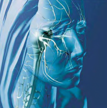
Pain from migraine headaches is typically located on only one side of the head, behind the eye.
Some researchers theorize that chronic headache sufferers lack normal levels of pain-blocking neurotransmitters called endorphins, a Greek word that means “the morphine within.” This deficiency means that their pain signals are more likely to cause severe discomfort than those in people who have higher endorphin levels.
Migraines are particularly devastating because of their severity and recurrence. They begin with impulses in hyperactive nerve cells. These impulses tell blood vessels in the head to constrict, and then to dilate. The process releases serotonin, prostaglandins, and other chemicals that inflame nerve cells surrounding the blood vessels in the brain. Specifically targeted are the trigeminal cranial nerve and its connections to the upper spinal cord and brain stem. The result: pain. Researchers long believed migraines arose from the narrowing and expanding of blood vessels on the surface of the brain; now, the most common theory traces migraines to hereditary abnormalities of the brain itself.
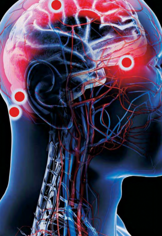
HEADACHES In the waning days of the Civil War, Union general Ulysses S. Grant was suffering from a terrible headache. He stopped at a farmhouse in the rear of his army, which had been pressing the forces of Confederate general Robert E. Lee. “I spent the night in bathing my feet in hot water and mustard, and putting mustard plasters on my wrists and the back part of my neck, hoping to be cured by morning,” Grant wrote in his journal on April 9, 1865.
Shortly afterward, Grant was visited by a messenger who carried a note saying Lee, who had refused to surrender the previous day, had changed his mind and would be willing to meet to discuss a formal end of hostilities. “When the officer reached me,” Grant said, “I was still suffering from the sick headache; but the instant I saw the contents of the note I was cured.”
Red indicates pain in a map of common headache sites, none of which is in the brain itself
Grant probably suffered from a muscle-contraction, or “tension,” headache. Typically, a tension headache begins when the neck, scalp, and face muscles are tensely held stiff for a long time. The most usual source is prolonged anxiety, a debilitating form of stress. Grant needed Lee to surrender; Lee’s announcement of his plans took the worries, and the agony, away. “The pain in my head seemed to leave me the moment I got Lee’s letter,” Grant reportedly told an aide as he rode off to end the war.
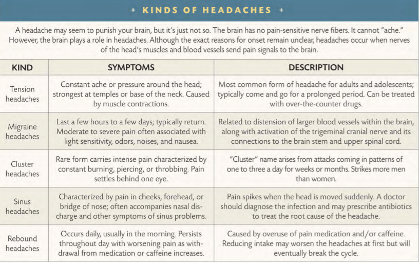
HEADACHES CATEGORIES
Even as it serves as an indicator that homeostasis is being disrupted, a headache is not a disease per se. Instead, it maya symptom of some other problem. It can manifest itself in response to irritation of blood vessels in the head, or to an injury or imbalance, or to inflammation of bodily tissues, to disorders related to stress-or to a host of other possible triggers. While it may feel as if the brain screams in pain, a headache can only occur outside the brain itself, which contains no pain receptors.
Headaches come in dozens of varieties. An easy way to categorize them is by the ways they cause pain. Muscle contractions such as Grant’s are one of the most common sources, especially among those living with high levels of stress. Dilation of blood vessels is a second typical cause. When arteries expand in the head, they squeeze against surrounding tissues, producing viselike pressure and pain. Fever, migraines, drug reactions, changes in blood pressure, and carbon dioxide poisoning can provoke dilation. Internal traction an abnormal growth in the head, for example is a third trigger. When a tumor presses against other tissues, or the brain itself begins to swell, the pressure causes pain. Inflammation is a fourth common source. Allergic reactions and infections such as meningitis can irritate pain-sensitive receptors in the head. Finally, headaches can occur without an obvious physical cause. These headaches are called psychogenic, meaning they arise in the psyche. They may spring from an emotional problem, as the sufferer converts emotional pain into real, physical symptoms.
The word migraine evolved from the Greek word hemikrania, meaning “half-skull.”
Many of these disorders strike not next to the brain, but in the eyes, sinuses, and other facial organs and tissues. Cranial nerves intimately connect the face and neck muscles to the brain, so it is no wonder pain sensations can spread until they feel as if they overwhelm the entire head.
Treating chronic headaches requires a proper diagnosis. Given the wide range of headaches and their causes, as well as the possibility of triggers working in combination, medical treatment often relies on detective work. At least, however, the efficacy of treatment has advanced since humanity first tried to cure a headache. A thousand years ago, Arabs recommended applying hot irons to the head, while a French medical treatise written in Latin urged sufferers to mix the brain of a vulture with oil and shove it up the nose. Today, modern pharmaceuticals, relax- ation techniques, and proper diet target dilation, tension, and other causes. One of the most effective pain relievers is common aspirin.


You must be logged in to post a comment.